Warning: mkdir(): Permission denied in /home/virtual/lib/view_data.php on line 81

Warning: fopen(upload/ip_log/ip_log_2024-11.txt): failed to open stream: No such file or directory in /home/virtual/lib/view_data.php on line 83

Warning: fwrite() expects parameter 1 to be resource, boolean given in /home/virtual/lib/view_data.php on line 84
Effect of Collagen Plug and Bone Graft on Bone Formation in Rabbit Extraction Model

J Korean Dent Assoc > Volume 62(10); 2024 > Article
Park, Song, Kim, Ryu, Jung, Kook, Oh, and Park: Effect of Collagen Plug and Bone Graft on Bone Formation in Rabbit Extraction Model

Abstract

1. Purpose

This study aimed to investigate the differences in bone regeneration capacity following extraction between a control group without biomaterial placement and experimental groups where biomaterials were inserted (including xenograft, synthetic bone-containing collagen plugs, and collagen plugs).

2. Materials and Methods

Bilateral extraction of maxillary first premolars or second premolars was performed, dividing the rabbits into four groups [① Group 1 :no bone graft or collagen plug inserted, ② Group 2: collagen plug inserted after extraction, ③ Group 3: collagen plug containing synthetic bone inserted after extraction, ④ Group 4: xenograft material inserted after extraction]. After euthanized the rabbit, Four and eight weeks postoperatively, the extent of bone formation were evaluated histologically and radiographically.

3 Results

The degree of new bone formation using histological analysis showed higher results in both the control groups at weeks 4 and 8, although not statistically significant. Across all groups, higher bone formation was observed at 8 weeks compared to 4 weeks. In radiological analysis using micro CT, Bone Mineral Density (BMD) and Bone Volume/Tissue Volume(BV/TV) were highest in Group 4. Trabecular bone thickness(Tb.Th) was highest in the control group, but not statistically significant.

4. Conclusion

In all groups, the degree of new bone formation at 4 and 8 weeks did not show statistically significant differences histologically and radiologically.

1. INTRODUCTION

Tooth extraction induces biological changes in the surrounding alveolar bone, resulting in subsequent alveolar bone resorption and irreversible vertical and horizontal ridge reduction [1]. Many studies have reported that approximately 30% of alveolar bone resorption occurs following extraction. Most bone resorption occurs within the first 6 months after extraction, with subsequent annual increases in resorption rates ranging from 0.5% to 1% [2]. Furthermore, most studies indicate that bone resorption occurs more extensively on the buccal aspect than on the lingual aspect. Reports show that the average vertical bone resorption is approximately 2mm greater on the buccal than the lingual side [3].
Araújo et al. showed that 29~63% of horizontal bone resorption and 11~22% of vertical bone resorption occurred within the first 6 months after tooth extraction [4]. Dental rehabilitation using implants is significantly affected by alveolar bone resorption.
Research on extraction socket preservation procedures has focused on preventing alveolar bone resorption following tooth extraction to reduce the necessity for ridge augmentation procedures. Extraction socket preservation refers to all procedures performed before and after tooth extraction to promote bone growth and prevent alveolar ridge resorption [5]. Extraction socket preservation helps reduce alveolar bone resorption and gingival recession, which can aid in preserving ideal bone height and volume for implant placement. Various bone graft substitutes, such as autogenous bone, allogeneic bone, xenogeneic bone, and synthetic bone, can be used in extraction socket preservation procedures. Autogenous bone grafting is considered the gold standard due to its osteogenic properties, although it has the disadvantages of high resorption rates and the need for secondary surgical sites. In contrast, allogeneic bone serves as a bone substitute with osteoinductive and osteoconductive properties, making it beneficial for clinical use.
Xenogeneic bone grafts, predominantly derived from bovine or porcine sources, are commonly used in dental procedures, with demineralized bovine bone being the most prevalent. Xenogeneic bone grafts provide osteoconductive properties and serve as scaffolds for bone regeneration. According to a meta-analysis by Canullo et al., xenogeneic bone grafts resulted in superior alveolar ridge preservation but histologically showed a lower degree of new bone formation.
A study by Piattelli et al. showed [6] that bovine bone grafts remained present even 10 years after ridge preservation. Therefore, xenogeneic bone grafts are considered non-resorbable biomaterials in current practice [7].
However, allografts and xenografts used as substitutes for autografts carry a rare but significant risk of transmitting various diseases, such as HIV, hepatitis, and prion-associated diseases. These drawbacks can be addressed by considering the transplantation of synthetic bone substitutes. These synthetic bone substitutes can be absorbable or non-absorbable and are known to possess osteoconductive properties similar to xenografts, although they lack osteoinductive capabilities. The osteoconductivity of these substitutes can vary depending on the composition of the graft material, manufacturing method, and physical characteristics [8]. Hydroxyapatite (HA) is known for its excellent biocompatibility because it is a major component of teeth and bones. HA offers strong mechanical properties with compressive strength exceeding 160 MPa [9]. However, HA is often combined with other bone substitutes rather than used alone. Among these, HA-collagen composites leverage the flexible nature of collagen to enhance HA's poor fracture toughness, offering advantages in durability. Beta-tricalcium phosphate(b-TCP) exhibits biocompatibility and biodegradability, possessing properties similar to the inorganic phase of bone. Complications, such as non-union and infection, are rarely reported in bone grafting using b-TCP. However, according to G. Fernandez de Grado et al., materials composed predominantly of TCP may have some limitations due to TCP's low physical resistance [10].
Absorbable collagen sponges can be inserted in addition to bone substitutes. Collagen sponges act as extracellular matrices within the socket, aiding osteoblast migration, stabilizing the blood clot, and assisting in connective tissue healing [11]. However, a study by Anderud J et al. reported no significant volumetric differences in alveolar bone changes between the control group without any intervention after extraction and the group with collagen plugs [12].
Collagen sponges containing synthetic bone substitutes, such as hydroxyapatite, tricalcium phosphate, or biphasic calcium phosphate with hyaluronic acid and gelatin, are sometimes used. T.B.L. Nguyen et al. reported successful fabrication of scaffolds composed of hyaluronic acid/gelatin and biphasic calcium phosphate for bone regeneration. Hyaluronic acid is crucial in promoting wound healing, while gelatin is widely used in medicine due to its excellent biodegradability [13].
Over the past 25 years, various treatment methods and graft materials in extraction and preservation procedures have been studied; however, none have been considered ideal [14,15]. Furthermore, according to Canullo et al., recent studies have investigated the self-regenerative capacity of bone defects occurring after extraction(without the placement of biomaterials) and the outcomes of healing following extraction [16]. Therefore, this study aimed to investigate the differences in bone regeneration capacity following extraction between a control group without biomaterial placement and experimental groups where biomaterials were inserted(including xenograft, synthetic bone-containing collagen plugs, and collagen plugs).

2. MATERIALS AND METHODS

2.1 Experimental materials

This study used eight New Zealand rabbits(aged 10~16 months, weighing 3.0~3.5kg, male). Anesthesia was induced using ketamine hydrochloride (60mg/kg) and xylazine hydrochloride(10mg/kg). Bovine bone devoid of proteins(Kisbone, Kisplant, Korea) was used as xenograft. Synthetic bone substitutes containing hydroxyapatite and β-tricalcium phosphate(B-TCP) were incorporated in collagen plugs(Qbonplug, Inobone, Korea). Collagen plugs derived from bovine sources were also used(Teruplug, Olympus Terumo Biomaterials Corp., Japan). Absorbable sutures were used(Vicryl 4.0, Ethicon, Inc., Raritan, NJ, USA). Postoperative analgesics and antibiotics administered were ketoprofen(3mg/kg) and gentamicin(4mg/kg), respectively. Euthanasia of the subjects was performed by intravenous injection of potassium chloride solution(1mg/kg) at 4 weeks and 8 weeks postoperatively.

2.2 Experimental method

The experiment was conducted with approval from the Chonnam National University Hospital Animal Care and Use Committee(CNUHCACUC-23017). Anesthesia was induced using ketamine hydrochloride(60mg/kg) and xylazine hydrochloride(10mg/kg), followed by infiltration anesthesia using 2% lidocaine with 1:100,000 epinephrine at the extraction site. Bilateral extraction of maxillary first premolars or second premolars was performed, dividing the rabbits into four groups [① Group 1(control): no bone graft or collagen plug inserted, ② Group 2: collagen plug inserted after extraction, ③ Group 3: collagen plug containing synthetic bone inserted after extraction, ④ Group 4: xenograft material inserted after extraction]. Groups were randomly assigned(at the 4th and 8th weeks, the sample size for each group is 2), and after socket preservation with bone graft or collagen plug insertion, closure was achieved with absorbable sutures. There were no complications, such as surgical site infections, and antibiotics and analgesics were administered by injection for three days postsurgery. Four and eight weeks after surgery, rabbits were euthanized by injecting potassium chloride solution intravenously to evaluate bone formation. Extraoral incisions were made to obtain the surgical sites, and the maxilla containing the experimental areas was excised using a saw(Figure 1).

2.3. Histologic analysis

2.3.1. Tissue processing and hematoxylin and eosin staining

After taking micro-CT, the specimens were dehydrated through a series of ethanol washes, followed by clearing in xylene according to standard protocols, and embedded in paraffin wax. The paraffinembedded sections were cut to a thickness of 5μm and stained with hematoxylin-eosin. Prepared samples were observed under an optical microscope, and images were captured using PANNORAMIC MIDI II(3DHistech Ltd, Hungary).

2.3.2. Histomorphometric analysis

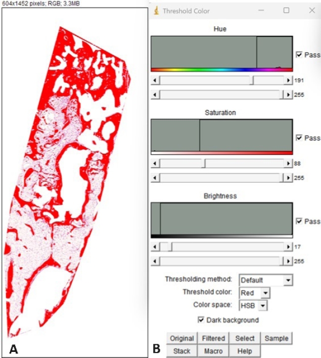
The ImageJ program(Wayne Rasband, National Institutes of Health, Bethesda, MD, USA) was used for a quantitative analysis of new bone formation. Following the method described by Jeremy R.,13 bone formation was evaluated by setting thresholds based on scanned tissue images, in which the region of interest was defined along the cortical border of the extraction site to avoid extension beyond the periodontal ligament(PDL) space of adjacent teeth, and saved in JPG format. A threshold color analysis tool within ImageJ was used to adjust the hue, saturation, and brightness values to select areas of bone formation. The measurement tool was then used to quantify the area filtered in red(representing bone formation)(Figure 2).

2.4. Radiological analysis using micro-CT

The excised maxilla was fixed in 4% paraformaldehyde solution and scanned using micro-CT(Scanco μ45, SCANCO Medical AG, Switzerland) under 90 kV and 88 μA with a 0.5 aluminum filter to assess the experimental sites.
Reconstruction was performed to exclude the periodontal ligament(PDL) space of adjacent teeth and cortical borders of the extraction socket. Bone mineral density(BMD), trabecular bone thickness(Tb. Th), and bone volume per total volume (BV/TV) were measured using micro-CT analysis software. BMD indicates the average hydroxyapatite(HA) amount within the bone, Tb.Th represents the width of bone structures, and BV/TV quantifies the amount of bone within the selected area(Figure 3).

2.5. Statistical analysis

Statistical analysis of the data was performed using IBM SPSS Statistics ver. 23.0 software(IBM Co., Armonk, NY, USA). Mann-Whitney and Kruskal-Wallis tests were used to determine significant differences among groups, with a statistical significance level set at p < 0.05.

3. RESULTS

3.1. Histologic analysis

3.1.1. Histological images of the experimental groups at 4 weeks after surgery

All groups showed new bone formation. The control group showed abundant osteoblasts surrounding the new bone and a rich extracellular matrix between the newly formed bone. Group 4, which underwent heterologous bone grafting, showed persistent graft material without degradation, surrounded by newly formed bone(Figure 4).
The group that received heterologous bone grafts was magnified by 5×. Newly formed bone was observed around the graft material, with abundant extracellular matrix(ECM) visible between them. Further magnification revealed osteoblasts lining the graft material and osteocytes within the newly formed bone(Figure 5).

3.1.2. Histological image of the experimental groups at 8 weeks after surgery

At the 4-week comparison, all groups exhibited lamellar bone formation at the central area adjacent to the extraction socket, and trabeculae appeared more uniformly straight. Additionally, in Group 4, the heterologous bone graft remained unabsorbed and observable, with increased surrounding newly formed bone compared to the 4-week assessment (Figure 6).

3.1.3 Histomorphometric analysis

The extent of bone formation within the extraction sockets was compared using Image J software. At 4 weeks, the groups showed the highest levels of new bone formation in the following order: control group, group with collagen plug containing synthetic bone, group with collagen plug alone, and group with heterologous bone graft. By 8 weeks, the order of highest new bone formation observed was: control group, group with heterologous bone graft, group with collagen plug containing synthetic bone, and group with collagen plug alone.
All groups showed higher levels of bone formation at 8 weeks compared to 4 weeks but with no statistically significant difference. The Kruskal-Wallis test was conducted to compare the degree of new bone formation among groups at each time point, showing no significant differences. Changes in bone formation between 4 and 8 weeks within each group compared using the Mann-Whitney U test yielded no significant results(Table 1).

3.2 Radiological analysis using micro-CT

BMD showed higher results in Group 4 at 4 and 8 weeks, while trabecular bone thickness was higher in the control group at both time points. BV/TV values also showed the highest results in Group 4. All three values of BMD, trabecular bone thickness, and BV/TV were higher at 8 weeks compared to 4 weeks in all groups but with no statistically significant results(Figure 7).

4. DISCUSSION

The healing process of the extraction socket occurs immediately after tooth extraction and continues for approximately six months. As the extraction socket remodeling progresses, inevitable crestal bone resorption and dimensional changes ensue. Adequate width and height of the alveolar ridge are crucial for implant restoration following extraction; insufficient dimensions necessitate compensatory procedures, such as ridge augmentation, alveolar ridge split procedures, and guided bone regeneration to facilitate implant placement. Recently, efforts have focused on minimizing post-extraction bone resorption by performing immediate socket preservation procedures following extraction. Research in this area is actively pursued to mitigate the need for additional surgical interventions [2,3,8,12,13,15,16,18,19,21].
Most currently used biomaterials for alveolar ridge preservation(ARP) do not accelerate bone healing or induce new bone formation in osseous defects compared to unassisted socket healing. Although the quantity of newly formed bone following ARP may be inferior to that achieved in unassisted socket healing, implant placement feasibility, osseointegration, survival and success rates, and susceptibility to peri-implant diseases are similar to those of implants placed in pristine bone. ARP may reduce the need for further, simultaneous bone augmentation at implant placement and can reduce the need for sinus augmentation in maxillary molars.
The dental alveolus is ideal for evaluating bone formation after tooth extraction due to certain factors present for graft material immobilization [17]. There are numerous studies conducted on calvarial defects for the advantage of creating multiple defects in a single subject. However, the surgical model of rabbit alveolus offers advantages in cost, space, storage, and maintenance [18].
All materials used for extraction socket preservation procedures in this study did not induce any inflammatory reactions; there was no statistically significant difference in the extent of new bone formation among all groups. Histologically analyzed, at both 4 and 8 weeks, the xenografts showed no signs of absorption; instead, new bone formation occurred around the non-absorbed xenografts. However, since there was no statistically significant difference in new bone formation compared to the control group, the presence of non-absorbed xenografts may not have influenced the extent of new bone formation.
According to Calixto et al. and Araufo et al. [19], specific particles or residual graft materials can delay bone regeneration. This delay is attributed to a foreign body reaction caused by the particles, with inflammatory cells observed histologically around the graft materials. Studies by Chan et al. and Covani et al. [20,21] suggest that socket preservation procedures may interfere with healing after extraction, offering no distinct advantages. They argue that various graft particles may remain indefinitely unabsorbed, potentially complicating subsequent autogenous bone replacement. Meta-analyses conducted by L. Canullo16 and others showed significantly lower levels of new bone formation in groups that underwent xenograft implantation compared to control groups without grafts after extraction. However, other studies suggest that non-absorbable bone graft materials may help maintain the volume of the alveolar ridge. Furthermore, while collagen plugs do not affect bone formation, they can reduce patient discomfort during the healing period after extraction.
Histological analysis revealed no inflammatory cells around the residual xenograft implants. Moreover, new bone formation was observed to a similar extent as in the control group, and the degree of new bone formation was highest in the control group compared to groups where bone grafts or collagen plugs were inserted, both at 4 and 8 weeks. Moreover, the amount of new bone increased over time, with all groups showing higher values at 8 weeks compared to 4 weeks. However, these differences were statistically insignificant. However, the limited sample size in this study renders the results susceptible to Type II errors. Although expanding the sample size is constrained by the nature of animal experimentation, increasing the number of specimens per animal—beyond the two specimens utilized in this study—could potentially alleviate these statistical limitations to some degree.
Covani et al. [22] mentioned the challenges of distinguishing between implanted bone particles and newly formed bone radiographically. They cautioned that radiographic measurements of the alveolar bone area may not accurately reflect the actual alveolar bone area, emphasizing the need for carefully interpreting experimental results related to bone studied solely through radiographic analysis. Therefore, this study used histological results in conjunction with radiographic analysis.
The BMD and BV/TV values analyzed through micro-CT imaging in this study did not entirely correlate with histological results. BMD and BV/TV values were higher in the groups with xenograft and collagen plug with synthetic bone inclusion compared to the control group, which could be attributed to the presence of particles from xenograft and synthetic bone, as mentioned by Covani et al. Therefore, particles from xenograft and synthetic bone may have influenced the amount of bone formation observed in micro-CT analysis. Thus, histological analysis with radiographic evaluation is essential in studies with bone grafts to accurately assess the amount of actual bone formation.
Victor F et al [23]. placed implants in the tibia of 28 rats divided into deproteinized bovine bone(DBB) and native bone(NB) groups. They reported higher osseointegration in the NB group than in the DBB group. Research on the impact of graft materials on implant osseointegration remains limited. Therefore, further research is needed to investigate whether there are differences in osseointegration in sites with bone grafting.
This study evaluated the extent of bone formation between experimental groups with collagen plug, collagen plug containing synthetic bone, and xenograft insertion compared to the control group histologically and radiographically. Although all groups showed new bone formation, statistically significant differences were absent. However, the ultimate purpose of bone grafting is for implant installation, raising the critical question of whether there is a difference in osseointegration between native and grafted bone during implant installation. There remains ongoing debate on implant success and survival rates following extraction and preservation procedures.

5. Conflicts of Interest

The researcher claims no conflicts of interest

Figure 1.
Excised maxilla
The maxilla containing the tooth extraction socket is resected using a saw.
jkda-2024-62-10-002f1.jpg
Figure 2.
Method for threshold color measurements (Image J Software)
A. Red area denotes the filtered region
B. Filtering is performed by adjusting hue, saturation, and brightness
jkda-2024-62-10-002f2.jpg
Figure 3.
Reconstruction of extraction socket using micro CT image
A. The region of interest (Red polygon) is defined to exclude the periodontal ligament (PDL) space and cortical border of adjacent teeth from the images acquired using scanco microCT.
B. Using the defined region of interest, the tooth extraction socket is reconstructed in 3D.
jkda-2024-62-10-002f3.jpg
Figure 4.
H&E images of the Control and experimental groups at 4 weeks postoperatively
(A,B,C,D = Scale bar is 2000 μm , A’,B’,C’,D’ = Scale bar is 500 μm)
A,A’: G1 (control: no bone graft or collagen plug inserted)
B,B’: G2 (Collagen plug collagen plug inserted after extraction)
C,C’: G3 (collagen plug containing synthetic bone inserted after extraction)
D,D’: G4 (bovine inserted after extraction)
jkda-2024-62-10-002f4.jpg
Figure 5.
H&E images of G4 (bovine inserted after extraction) at 4 weeks postoperatively
A: Scale bar is 500 μm, B: An image zoomed in at 5x magnifiaction of the rectangular area in Figure
A (Scale bar is 100 μm) Osteoblastic reaming (blue arrow), osteocyte in the new bone (black arrow),GB: Grafted bone, NB: New bone
jkda-2024-62-10-002f5.jpg
Figure 6.
H&E images of the Control and experimental groups at 8 weeks postoperatively
(A,B,C,D = Scale bar is 2000μm , A’,B’,C’,D’ = Scale bar is 500μm
A,A’: G1 (control: no bone graft or collagen plug inserted)
B,B’: G2 (Collagen plug collagen plug inserted after extraction)
C,C’: G3 (collagen plug containing synthetic bone inserted after extraction)
D,D’: G4 (bovine bone inserted after extraction)
jkda-2024-62-10-002f6.jpg
Figure 7.
The values of BMD, Tb.Th, and BV/TV at 4 and 8 weeks postoperatively in each group [G1: Control, G2: Collagen plug, G3: Collagen plug including synthetic bone, G4: Bovine bone]
jkda-2024-62-10-002f7.jpg
Table 1.
Bone volume fraction in extraction socket at 4 and 8 weeks postoperatively in each group
Group Material Bone volume fraction (%) (mean ± SD)
4 weeks 8 weeks
1 None 41.2 ± 8.5 45.2 ± 2.3
2 Collagen plug 37.1 ± 0.4 38.8 ± 0.4
3 Collagen plug including synthetic bone 38.3 ± 8.8 38.9 ± 0.3
4 Bovine bone 35.7 ± 6.8 40.2 ± 2.3

REFERENCES

1. Schropp L., Wenzel A., Kostopoulos L., Karring T.. Bone Healing and Soft Tissue Contour Changes Following Single-Tooth Extraction: A Clinical and Radiographic 12-Month Prospective Study. Int. J. Periodontics Restor. Dent 2003;23:313–323.
2. Lin HK, Pan YH, Salamanca E, Lin YT, Chang WJ. Prevention of Bone Resorption by HA/β-TCP + Collagen Composite after Tooth Extraction: A Case Series. Int J Environ Res Public Health 2019;Nov 21 16(23):4616.
crossref pmid pmc
3. Araújo M.G., Lindhe J.. Dimensional Ridge Alterations Following Tooth Extraction. An Experimental Study in the Dog. J. Clin. Periodontol 2005;32:212–218.
crossref pmid
4. Araújo M.G., Sukekava F., Wennström J.L., Lindhe J.. Ridge Alterations Following Implant Placement in Fresh Extraction Sockets: An Experimental Study in the Dog. J. Clin. Periodontol 2005;32:645–652.
crossref
5. Matej Tomas et al. A Histologic, Histomorphometric, and Immunohistochemical Evaluation of Anorganic Bovine Bone and Injectable Biphasic Calcium Phosphate in Humans: A Randomized Clinical Trial. Int. J. Mol. Sci 2023;24:5539.
crossref pmid
6. Kim Y., Nowzari H., Rich S.K.. Risk of Prion Disease Transmission through Bovine-Derived Bone Substitutes: A Systematic Review. Clin. Implant. Dent. Relat. Res 2013;15:645–653.
crossref pmid
7. Piattelli M., Favero G., Scarano A., Orsini G., Piattelli A.. Bone Reactions to Anorganic Bovine Bone (Bio-Oss) Used in Sinus Augmentation Procedures: A Histologic Long-Term Report of 20 Cases in Humans. Int. J. Oral Maxillofac. Implant 1999;14:835–840.
8. Fukuba S, Okada M, Nohara K, Iwata T. Alloplastic Bone Substitutes for Periodontal and Bone Regeneration in Dentistry: Current Status and Prospects. Materials. 2021;14(5):1096.
9. Koshino T, Murase T, Takagi T et al. New bone forma tion around porous hydroxyapatite wedge implanted in opening wedge high tibial osteotomy in patients with oste oarthritis. Biomaterials 2001;22:1579–1582.
crossref
10. Fernandez de Grado G, Keller L, Idoux-Gillet Y, Wagner Q, Musset A.-M, Benkirane-Jessel N, Bornert F, Offner D. Bone substitutes: a review of their characteristics, clinical use, and perspectives for large bone defects management. J. Tissue Eng 9:2018;2041731418776819.
pmid pmc
11. Konishi J, Miyamoto T, Miura A, Osaki K. An investigation into the wound healing action of the atelocollagen tooth extraction wound protection material (TRE-641) on tooth extraction site. J Jpn Soc Biomater 1998;16:266–75.
12. Anderud J, Lennholm C, WälivaaraWaiver DÅ. Ridge preservation using Collacone compared with an empty socket: a pilot study. Oral Surg Oral Med Oral Pathol Oral Radiol 2021;Aug;132(2):e55–e61. 21;16(23):4616.
crossref pmid
13. Nguyen B.L., Lee B.-T.. A combination of biphasic calcium phosphate scaffold with hyaluronic acid-gelatin hydrogel as a new tool for bone regeneration. Tissue Eng A 20 (13-14):2014;1993–2004.
crossref
14. Tan-Chu JH, Tuminelli FJ, Kurtz KS, Tarnow DP. 2014;Analy sis of buccolingual dimensional changes of the extraction socket using the “ice cream cone” flapless grafting technique. Int J Periodontics Restorative Dent 34(3):399–403.
crossref pmid
15. Atieh MA, Alsabeeha NH, Payne AG, Ali S, Faggion CMJ, Esposito M. 2021;Interventions for replacing missing teeth: alveolar ridge preservation techniques for dental implant site development. Cochrane Database Syst Rev 4:CD010176.
crossref pmid
16. Canullo L., Del Fabbro M., Khijmatgar S. et al. Dimensional and histomorphometric evaluation of biomaterials used for alveolar ridge preservation: a systematic review and network meta-analysis. Clin Oral Invest 26:141–158. 2022.
crossref pdf
17. Becker W, Becker BE, Caffesse R. A comparison of demineralized freeze-dried bone and autologous bone to induce bone formation in human extraction sockets. J Periodontol 1994;Dec;65(12):1128–33.
crossref pmid pdf
18. Cantin M. Alveolar Ridge conservation by early bone formation after tooth extraction in rabbits: a histomorphological study. International Journal of Morphology 33(1):369–374.
crossref
19. Calixto RF, Teófilo JM, Brentegani LG, Lamano-Carvalho TL. Grafting of tooth extraction socket with inorganic bovine bone or bioactive glass particles: comparative histometric study in rats. Implant Dent 2007;Sep;16(3):260–9.
crossref pmid
20. Chan H.L., Lin G.H., Fu J.H., Wang H.L.. Alterations in bone quality after socket preservation with grafting materials: A systematic review. Int. J. Oral Maxillofac. Implant 2013;28:710–720.
crossref
21. Covani U., Giammarinaro E., Marconcini S.. A New Approach for Lateral Sinus Floor Elevation. J. Craniofac. Surg 2020;31:2320–2323.
crossref pmid
22. Covani U, Giammarinaro E, Panetta D, Salvadori PA, Cosola S, Marconcini S. Alveolar Bone Remodeling with or without Collagen Filling of the Extraction Socket: A High-Resolution X-ray Tomography Animal Study. Journal of Clinical Medicine 2022;11(9):2493.
crossref pmid pmc
23. Quiroz VF, Lima JR, Pinotti FE, Marcantonio RA, Marcantonio E, Oliveira GJ. Comparison of osseointegration in areas grafted with deproteinized bovine bone and native bone. A preclinical study. Acta Odontol Latinoam 2022;Apr 30 35(1):3–9.
crossref pmid pmc


Editorial Office
Korean Dental Association, 257 Gwangnaru-ro, Seongdong-gu, Seoul 04802, Korea
Tel: +82-2-2024-9100   Fax: +82-2-468-4655/58   

Copyright © 2024 by Korean Dental Association.

Developed in M2PI