Odontogenic deep neck infection progressing to descending necrotizing mediastinitis and sepsis: A case report
Article information

Abstract
Descending necrotizing mediastinitis (DNM) is a rare but potentially fatal complication of odontogenic infection. A 62-year-old woman with diabetes mellitus presented with left facial swelling and sepsis manifested by severe hypotension and hypoxemia; the source was a periapical abscess of the left mandibular second premolar. Contrast-enhanced CT showed a deep neck abscess with gas-containing collections extending along the left carotid space into the mediastinum, accompanied by marked airway narrowing. Emergency cervical incision and drainage confirmed necrotizing fasciitis. On post-admission day 8, follow-up CT demonstrated progression of the mediastinal gas locules to a frank abscess requiring thoracic surgical drainage. This case illustrates how a localized odontogenic infection can progress to DNM and highlights serial contrast-enhanced CT as an adjunct for assessing disease progression and the need for additional surgical source control. Successful management requires airway control, broad-spectrum antimicrobial therapy, adequate cervical and mediastinal drainage, intensive sepsis care, and multidisciplinary collaboration.
Introduction
Odontogenic infections usually arise from periapical lesions, periodontal disease, or pericoronitis, and although most remain localized, a subset can progress along fascial planes into the deep cervical spaces and mediastinum, making early recognition critically important [1,2]. Odontogenic deep neck infections may lead to life-threatening complications when diagnosis or drainage is delayed, particularly when infection extends into the retropharyngeal space or mediastinum [3-5]. For clarity, a deep neck abscess refers to a localized purulent collection within the deep cervical fascial spaces, whereas cervical necrotizing fasciitis refers to a rapidly progressive infection characterized by necrosis of the cervical fascia and adjacent soft tissues. Descending necrotizing mediastinitis (DNM) refers to the descending extension of a severe cervical or oropharyngeal infection into the mediastinum along the deep fascial planes. Estrera et al. established diagnostic criteria based on clinical, radiographic, and operative findings [6]. Descending mediastinal spread may be associated with airway compromise, pleural or pericardial involvement, and poor clinical outcomes [4,7]. In odontogenic DNM, sepsis should be recognized as a systemic, life-threatening manifestation of severe infection that may arise concurrently with, rather than solely as a late complication of, mediastinal extension. Defined as life-threatening organ dysfunction caused by a dysregulated host response to infection, sepsis requires urgent recognition, hemodynamic resuscitation, early antimicrobial therapy, and prompt surgical source control [8-11]. Despite advances in imaging, antibiotic therapy, and surgical drainage, odontogenic DNM remains a rare but potentially fatal condition requiring prompt multidisciplinary management [5,11,12]. Contrastenhanced CT plays a central role in early diagnosis and surgical planning, as clinical findings may be nonspecific before advanced disease becomes apparent [3-5]. This report describes a case of odontogenic DNM complicated by an extensive deep neck abscess, a mediastinal abscess, airway compromise, and sepsis requiring intensive care. This case emphasizes the role of contrast-enhanced CT as an adjunct to serial clinical assessment and the need for coordinated airway management, antimicrobial therapy, surgical source control, and sepsis care within a multidisciplinary treatment strategy.
Case Report
This case report was reviewed by the Institutional Review Board of Chosun University Dental Hospital (CUDHIRB]2505-004). It was determined to be exempt from full review, as it involved a retrospective analysis of anonymized patient data.
A 62-year-old female patient visited the Department of Oral and Maxillofacial Surgery at Chosun University Dental Hospital with the chief complaint of left facial swelling. The patient reported that the onset of left facial swelling occurred 9 days before presentation. Two days before presentation, she underwent incision and drainage and received intravenous antibiotics at a local clinic; however, her symptoms persisted without improvement. At presentation, she complained of severe pain in the left facial and cervical regions, dysphagia, odynophagia, and dyspnea. The chronological clinical course is summarized in Table 2.
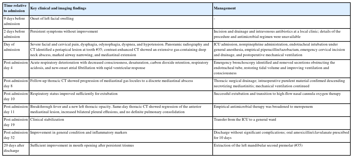
Chronological summary of the clinical course from symptom onset to definitive management of the odontogenic source.
The patient had underlying type 2 diabetes mellitus and hyperlipidemia. She had been unable to take her usual oral antihyperglycemic medications for approximately 1 week before admission because of progressive dysphagia and odynophagia. On presentation, her body temperature was 36.5°C, heart rate was 85 beats/min, respiratory rate was 21 breaths/min, and blood pressure was 80/40 mmHg. Despite the absence of fever, the patient showed marked hypotension and an elevated respiratory rate consistent with the reported dyspnea, suggesting systemic compromise associated with severe infection. Initial laboratory findings revealed marked leukocytosis with neutrophil predominance and elevated inflammatory markers. The white blood cell count was 23.41 × 10³/μL, with 89.6% neutrophils. High-sensitivity C-reactive protein and procalcitonin levels were markedly elevated to 39.6 mg/dL and 2.87 ng/mL, respectively. Arterial blood gas analysis showed severe hypoxemia, with a pO₂ of 33.4 mmHg and an oxygen saturation of 60.9%. Although the platelet count was preserved, coagulation studies demonstrated prolonged prothrombin time, elevated PTINR, and increased D-dimer and FDP levels, suggesting activation of the coagulation and fibrinolytic systems. A glycated hemoglobin level of 7.7% suggested suboptimal chronic glycemic control, although the admission glucose level was 114 mg/dL. No chronic diabetic complications were documented in the available medical records, and renal function was preserved, with a serum creatinine level of 0.84 mg/dL and an estimated glomerular filtration rate of 79 mL/min/1.73 m² on admission. These findings supported the clinical diagnosis of sepsis under the Sepsis-3 framework, given the presence of severe odontogenic infection with systemic organ dysfunction, including vasopressor-requiring hypotension and severe hypoxemia (Table 1). On physical examination, diffuse swelling with local heat, tenderness, and induration was noted over the left submandibular and upper cervical regions. Mouth opening was severely limited (trismus), with a maximal interincisal opening (MIO) of 10 mm, which precluded adequate intraoral examination.
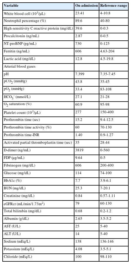
Laboratory results on admission.
Because intraoral examination was limited, the odontogenic source was identified radiographically. A panoramic radiograph revealed periapical radiolucency associated with the left mandibular second premolar (#35), accompanied by severe coronal caries. This finding was confirmed by CT images obtained with bone window settings, which demonstrated a periapical lesion of the same tooth (Fig. 1). Contrast-enhanced CT demonstrated extensive abscess formation with multiple gas locules involving the left facial and deep neck spaces, accompanied by diffuse soft tissue swelling and marked upper airway narrowing (Fig. 2). Gas-containing collections extended inferiorly along the left carotid space, suggesting that the carotid space served as the principal route of descending spread. The adjacent left internal jugular vein showed slit-like narrowing due to severe extrinsic compression, reflecting the mass effect of the surrounding inflammatory process. Gas locules were also traceable from the infected deep neck spaces into the visceral compartment of the mediastinum at presentation (Fig. 2I), supporting early mediastinal extension despite prior incision and drainage at a local clinic two days earlier. Coronal reformations demonstrated inferior extension of the infection toward the mediastinum with associated airway deviation (Fig. 3A and B). Sagittal reformations revealed abscess formation within the retropharyngeal space (Fig. 3C and D), raising particular concern because of its close anatomical relationship to the airway and its potential contribution to airway compromise. Based on these imaging findings, the patient was diagnosed with severe odontogenic deep neck infection with imaging findings suspicious for DNM. Thus, at the time of presentation, the patient had concurrent sepsis and suspected descending necrotizing mediastinitis, two life-threatening conditions that required immediate multidisciplinary intervention.
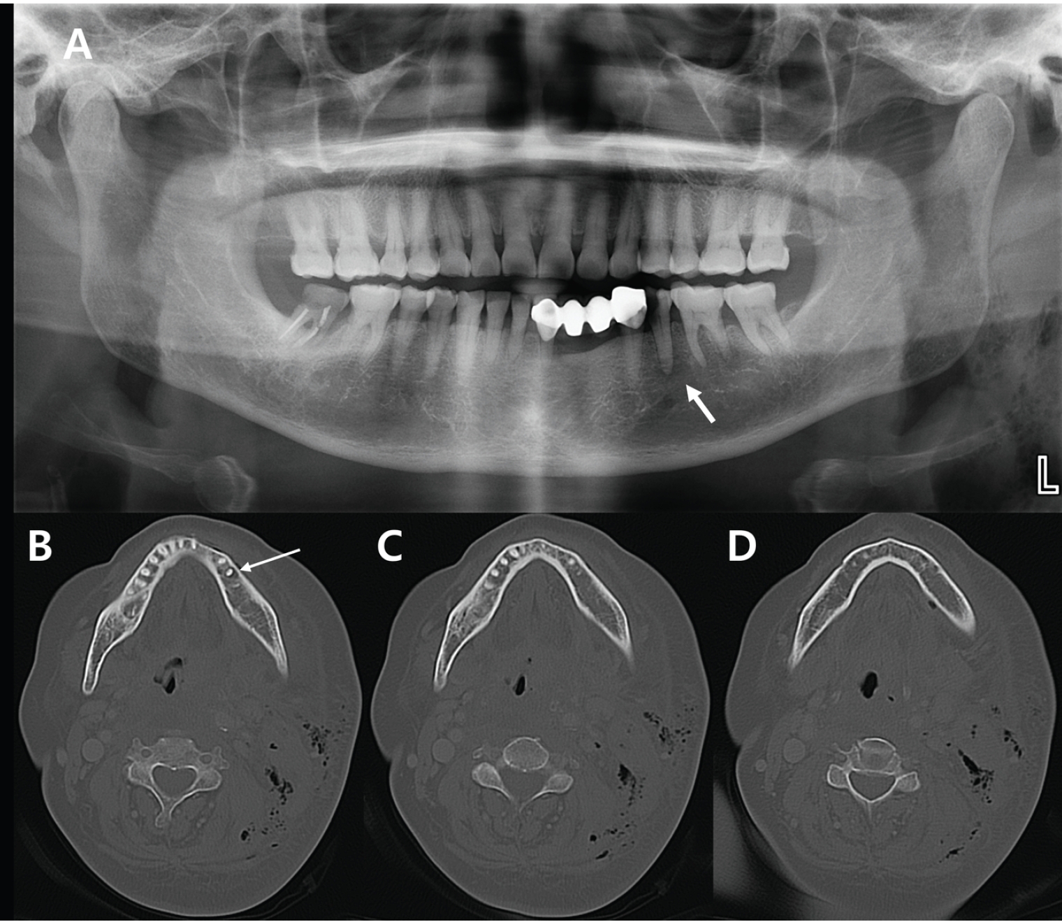
A. A panoramic radiograph shows a diffuse, non-corticated periapical radiolucency associated with the left mandibular second premolar (#35), accompanied by severe dental caries, suggestive of a periapical abscess. Multiple air bubbles are also observed in the soft tissues of the left neck. B-D. CT images with bone window settings demonstrate a periapical lesion involving the left mandibular second premolar (#35), with air bubbles also visible in the adjacent soft tissue of the left neck and mandibular region.
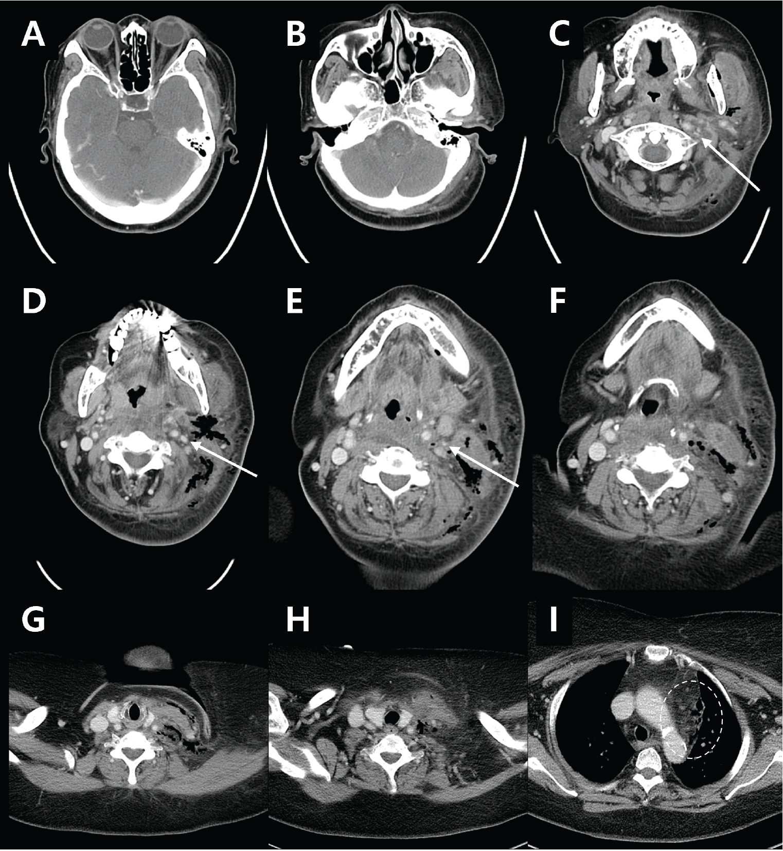
Initial contrast-enhanced CT images obtained at admission demonstrate extensive abscess formation with multiple air bubbles along the left deep neck spaces, accompanied by diffuse soft tissue edema and swelling. The marked soft tissue swelling results in significant narrowing of the airway. A and B. Abscess formation is observed in the left masticator space. C-H. Gas-forming abscess collections extend along the left carotid space, consistent with the carotid space as the anatomical route of inferior spread. The adjacent left internal jugular vein demonstrates a slit-like appearance due to severe extrinsic compression, secondary to the inflammatory mass effect of cervical necrotizing fasciitis (arrows). I. Gas locules extend inferiorly from the infected left deep neck spaces into the visceral compartment of the mediastinum, consistent with descending mediastinal spread of infection (dashed circle).
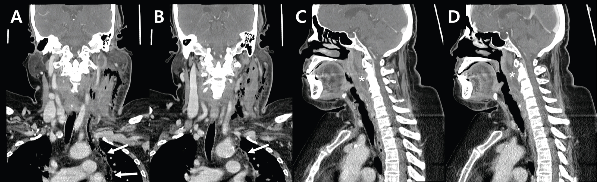
Coronal and sagittal contrast-enhanced CT images obtained at admission. A and B. Coronal images demonstrate extension of the left deep neck infection into the mediastinum (arrows) and associated airway deviation. C and D. Sagittal images demonstrate abscess formation within the retropharyngeal space (asterisks).
The patient was hospitalized and transferred to the intensive care unit (ICU) for close monitoring and management. Because of persistent hypotension, norepinephrine was administered, after which her blood pressure was maintained within the normal range. During the critical phase of illness, the patient’s oral antihyperglycemic medications were withheld, and glycemic control was managed with basal insulin glargine and rapid-acting insulin aspart, with dose adjustment according to enteral feeding. Glucose levels intermittently exceeded 200 mg/dL during hospitalization, with a peak value of 245 mg/dL. On the day of admission, the airway was secured with endotracheal intubation for general anesthesia, and emergency incision and drainage of the left deep neck abscess was collaboratively performed by the Departments of Otorhinolaryngology and Oral and Maxillofacial Surgery. Purulent discharge was obtained during the procedure, and intraoperative findings of necrotic tissue confirmed the diagnosis of necrotizing fasciitis. Postoperative mechanical ventilation was continued in the ICU. During the initial cervical drainage, closed- and open-pus specimens obtained from the left neck were submitted for Gram staining, bacterial culture, and antimicrobial susceptibility testing. Gram stains demonstrated gram-positive cocci together with gram-negative and gram-positive rods, and Streptococcus anginosus was isolated from both specimens. The open-pus isolate was susceptible to benzylpenicillin, ampicillin, cefotaxime, ceftriaxone, erythromycin, clindamycin, tetracycline, vancomycin, levofloxacin, linezolid, and tigecycline, with intermediate susceptibility to chloramphenicol. The closed-pus isolate was susceptible to penicillin, ampicillin, chloramphenicol, ciprofloxacin, erythromycin, clindamycin, tetracycline, and vancomycin, but resistant to gentamicin. Blood cultures obtained at admission and repeated two days later showed no growth. Empirical piperacillin/tazobactam was initiated on admission. Antimicrobial management was conducted in consultation with the Department of Infectious Diseases. Following two initial doses on the first day, treatment was continued at 4.5 g intravenously every 6 hours for 10 days.
On post-admission day 4, the patient developed acute respiratory deterioration with a decreased level of consciousness, desaturation, carbon dioxide retention, and new-onset atrial fibrillation with rapid ventricular response. Emergency bronchoscopy revealed obstruction of the endotracheal tube by secretions, and bronchoscopic removal restored tidal volume, with subsequent improvement in carbon dioxide retention and respiratory acidosis and recovery of alertness.
On post-admission day 8, comparison of the follow-up thoracic CT with the admission study demonstrated interval progression of the mediastinal gas locules to a frank abscess within the left aspect of the visceral compartment of the mediastinum (Fig. 4). Because this discrete mediastinal collection represented an anatomically distinct focus requiring additional thoracic source control, thoracic surgical drainage was performed. Purulent material was identified within the mediastinum during surgery, providing operative confirmation of DNM. A thoracic drainage specimen obtained during the procedure showed no organisms on Gram staining and no growth on routine bacterial culture; acid-fast bacillus testing was also negative. Mechanical ventilation was continued after thoracic drainage. On post-admission day 10, the patient was successfully extubated and transitioned to high-flow nasal cannula oxygen therapy. On post-admission day 11, she developed breakthrough fever, and serial chest radiographs demonstrated a newly developed left thoracic opacity, initially raising concern for hospital-acquired or aspiration pneumonia. Antimicrobial therapy was therefore empirically broadened to meropenem at 2 g intravenously every 8 hours. This change was based on the clinical and chest radiographic findings rather than antimicrobial resistance of the S. anginosus isolates or deterioration in renal function. Thoracic CT performed on the same day demonstrated interval regression of the anterior mediastinal lesion and increased bilateral pleural effusions, without definite pulmonary parenchymal consolidation. Meropenem was administered at 2 g intravenously every 8 hours for 21 days, followed by oral amoxicillin/clavulanate 625 mg three times daily for 10 days after discharge. Following clinical stabilization, the patient was transferred from the intensive care unit to a general ward on post-admission day 19. After infection control, the patient was switched from insulin back to her usual oral antihyperglycemic medications before discharge. Her general condition and inflammatory markers gradually improved, and she was discharged 32 days af-ter admission without significant complications. Because persistent trismus precluded immediate extraction, definitive treatment of the left mandibular second premolar (#35), considered the presumed odontogenic source, was deferred. After sufficient improvement in mouth opening, the tooth was extracted 20 days after discharge.
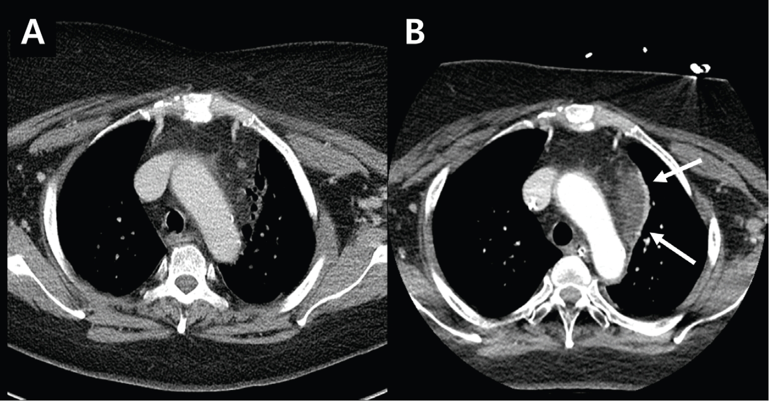
Comparative contrast-enhanced CT images demonstrating interval change in the left aspect of the visceral compartment of the mediastinum between admission (A) and post-admission day 8 (B). The gas bubbles previously identified at admission have progressed to frank abscess formation.
Discussion
This case illustrates the potential for an odontogenic infection to progress from a localized periapical lesion to severe multispace deep neck infection, descending necrotizing mediastinitis, airway compromise, and sepsis requiring intensive care. The present case showed several features associated with a severe clinical course, including diabetes mellitus, marked leukocytosis, extensive multispace involvement, and gas formation, several of which have been linked to life-threatening complications, ICU admission, and repeated surgery in odontogenic deep neck infections [2,13,14].
The route of spread was anatomically consistent with descending extension through the deep cervical fascial spaces. DNM has traditionally been diagnosed according to the criteria proposed by Estrera et al., including severe cervical infection, characteristic radiographic findings, operative or postmortem evidence of necrotizing mediastinal infection, and continuity between the cervical and mediastinal processes [6]. CT demonstrated gas-containing collections extending inferiorly along the left carotid space toward the superior mediastinum. Follow-up CT demonstrated progression of the initial mediastinal gas locules to a discrete abscess, prompting additional thoracic surgical drainage. Although the retropharyngeal– retrovisceral pathway is more commonly emphasized, the carotid space is a recognized anatomical route for cervicomediastinal spread [4,5].
Contrast-enhanced CT played an important adjunctive role in diagnosis and therapeutic decision-making by delineating the periapical source, gas-containing abscesses, airway narrowing, and mediastinal extension [4,15]. A previous report also emphasized the value of contrastenhanced CT in assessing deep neck infection with necrotizing fasciitis, airway compromise, and the need for repeated drainage [16]. The interval progression from mediastinal gas locules at admission to a discrete abscess on post-admission day 8 demonstrates that serial CT, when interpreted together with the clinical course, can identify newly drainable collections and support decisions regarding additional surgical source control [1,3,5].
Emergency cervical incision and drainage were performed on the day of admission, and intraoperative findings of necrotic tissue confirmed cervical necrotizing fasciitis. However, the initial cervical drainage did not eliminate the need for further surveillance, as the mediastinal component evolved into a drainable abscess on follow-up CT, necessitating additional thoracic surgical drainage. This staged approach is consistent with reports emphasizing adequate drainage of all infected compartments alongside broad-spectrum antimicrobial therapy and intensive care support [5,15]. Definitive management of the odontogenic source is an essential component of treatment. Early removal of the causative tooth, when clinically feasible, has been associated with a shorter hospital stay in patients with odontogenic deep neck infections [2]. In the present case, extraction of the left mandibular second premolar (#35) was deferred because persistent trismus precluded immediate dental treatment. After the acute deep neck and mediastinal infections had been controlled and mouth opening had sufficiently improved, the tooth was extracted 20 days after discharge as definitive treatment of the presumed odontogenic source. This sequence underscores the importance of completing treatment of the causative tooth as soon as the patient’s systemic and local conditions permit.
Severe odontogenic deep neck infections and DNM are typically polymicrobial, involving facultative or microaerophilic oral streptococci together with obligate anaerobic organisms [1,3,17,18]. The anaerobic component commonly includes gram-positive anaerobic cocci and gram-negative rods such as Prevotella, Porphyromonas, and Fusobacterium species [18]. Facultative streptococci may predominate during the early cellulitic phase and facilitate the spread of infection, whereas anaerobic organisms are particularly important during subsequent suppuration and abscess formation; anaerobic gramnegative rods have also been associated with necrotic abscesses [18]. In mixed infections, oxygen consumption by facultative organisms may create a reduced environment favorable for anaerobic growth, and synergistic interactions among bacterial species may increase the pathogenicity of the infecting flora [18]. In DNM, gram-positive cocci and gram-negative rods such as Fusobacterium have been associated with gas production and tissue injury [3,4]. These microbiological characteristics may partly explain the rapid progression of severe odontogenic infection through the cervical fascial spaces. Although the gas locules helped delineate the extent of infection and the route of descending spread, they were not specific for a particular gas-producing pathogen, particularly because some of the cervical gas could have represented procedural air introduced during the pre-referral incision and drainage.
In the present case, Streptococcus anginosus was isolated from both cervical pus specimens and was broadly susceptible to the tested β-lactam agents and clindamycin. However, multiple bacterial morphotypes were observed on Gram staining, and the patient had received antimicrobial treatment before referral. Therefore, recovery of S. anginosus alone may not have represented the complete microbial composition, and involvement of anaerobic copathogens that were not recovered by culture could not be excluded. Piperacillin/tazobactam was initially administered to provide broad empirical coverage of both oral streptococci and anaerobic organisms. This approach was consistent with the polymicrobial nature of severe odontogenic infections, as the tazobactam component extends the activity of piperacillin against many β-lactamase–producing anaerobic gram-negative rods [18]. The susceptibility results confirmed activity against the recovered S. anginosus isolates, whereas the possibility of undetected anaerobic copathogens supported continued broad-spectrum coverage during the acute phase. The subsequent escalation to meropenem was not prompted by antimicrobial resistance or worsening renal function, but was an empirical response to breakthrough fever and a newly developed left thoracic opacity that initially raised concern for hospital-acquired or aspiration pneumonia. Although microbiological testing was performed, complete characterization of the infecting microbiota remained limited by prior antimicrobial exposure and the failure to recover anaerobic organisms from culture.
The progression in this patient was likely multifactorial rather than attributable to the host systemic condition alone. The 9-day interval from symptom onset to tertiary evaluation may have permitted further spread through the deep cervical spaces before definitive imaging and multidisciplinary treatment [5,7,11]. Although incision and drainage had been performed and intravenous antibiotics had been administered before referral, details of the procedure and antimicrobial regimen were unavailable; therefore, the adequacy of the initial drainage and antimicrobial coverage could not be determined. At the time of referral, persistent infection and an untreated odontogenic source indicated that complete source control had not yet been achieved. The microbiological findings discussed above suggest that bacterial virulence, mixed microbial composition, and possible aerobic–anaerobic synergy may also have contributed to abscess formation and fascial spread [3,18]. In contrast, the inpatient empirical regimen, as described above, provided appropriate coverage for the recovered organism and suspected anaerobic copathogens, making inadequate antimicrobial coverage at the tertiary center an unlikely principal cause of progression. Thoracic drainage was performed when serial CT demonstrated a discrete mediastinal abscess on post-admission day 8. Although the admission CT had already shown mediastinal gas, a discrete mediastinal abscess became evident only on follow-up imaging. This case therefore underscores the importance of early thoracic surgical involvement, close serial reassessment, and prompt drainage when a mediastinal collection becomes apparent [5,15,17].
Sepsis represented an equally important systemic and life-threatening aspect of this case. Under the Sepsis-3 framework, sepsis is defined as life-threatening organ dysfunction caused by a dysregulated host response to infection, and clinical criteria such as SOFA and qSOFA were developed to identify patients at risk of poor outcomes [8,19]. Neither a SOFA score nor a qSOFA score was prospectively recorded, and retrospective scoring was limited because the level of consciousness at presentation was not documented in a form suitable for scoring. Sepsis severity was therefore characterized by the documented organ dysfunction—vasopressor-requiring hypotension and severe hypoxemia—rather than by a reconstructed score, supporting the clinical diagnosis of sepsis. Notably, the absence of fever did not reduce the severity of the systemic presentation, as sepsis may manifest primarily through organ dysfunction rather than pyrexia. The updated septic shock criteria further emphasize that vasopressor-dependent hypotension combined with elevated lactate identifies a particularly high-risk subset of sepsis, underscoring the importance of carefully distinguishing sepsis from septic shock in patients with circulatory compromise [20]. Early airway control and continued airway surveillance, ICU transfer, norepinephrine administration, broad-spectrum antimicrobial therapy, and staged cervical and mediastinal drainage were coordinated components of multidisciplinary management rather than independent interventions [5,9,10,15].
In conclusion, odontogenic infection can progress rapidly to descending necrotizing mediastinitis, airway compromise, and sepsis, even when the initial source appears localized. Serial contrast-enhanced CT should be regarded as an important adjunct for assessing disease progression and identifying the need for additional surgical source control, rather than as a stand-alone determinant of treatment success. Effective management requires early diagnosis, airway control and continuous airway surveillance, prompt broad-spectrum antimicrobial therapy, adequate drainage of cervical and mediastinal infection, intensive sepsis care, and coordinated multidisciplinary collaboration.
Notes
Conflicts of Interest
None