Biofluorescence-guided oral care for hospitalized geriatric patient with pneumonia: A case report
Article information

Abstract
Effective detection and removal of pathogenic oral biofilm in hospitalized geriatric patient are important for preventing aspiration pneumonia. This case report describes the clinical application of biofluorescence-based technology for immediate assessment of oral hygiene status and biofluorescence-guided oral hygiene care in a hospitalized geriatric patient with pneumonia. The thickness and extent of oral biofilm were difficult to determine by visual inspection; however, biofluorescence imaging revealed red biofluorescence across multiple regions of the oral cavity. Based on these findings, mechanical and chemical biofilm control was performed at the bedside by a dental professional. Following oral care, the red biofluorescence observed before intervention was no longer present, allowing objective evaluation of biofilm removal. These findings suggest that biofluorescence enables real-time visualization of pathogenic biofilm and objective monitoring of changes in oral hygiene, improving the objectivity of oral hygiene assessment and supporting quality improvement in oral care for hospitalized patients.
Introduction
Pneumonia in older adults is a serious disease associated with high hospitalization rates, morbidity, and mortality [1]. Hospitalized patients are particularly vulnerable to deterioration in oral hygiene due to high care dependency, limited access to dental services, and difficulties in maintaining self-performed oral care [2]. Impaired oral functions such as dysphagia or reduced salivary clearance can further promote oral biofilm accumulation and the formation of membranous substances on the oral mucosa [3]. Oral biofilm may serve as a reservoir of respiratory pathogens that can be aspirated into the lower airway, contributing to pneumonia development in vulnerable individuals [4,5]. Professional oral care has been shown to reduce pneumonia in immunocompromised older adults [4,6]; therefore, accurate identification and control of pathogenic oral biofilm are essential in this population.
Plaque-disclosing agents are widely used for visualization; however, their use may be limited by procedural complexity, soft-tissue staining, patient discomfort, and aspiration risk in bedridden patients [7]. Quantitative light-induced fluorescence (QLF) technology enables visualization and quantification of dental biofilm maturity based on biofluorescence [8]. QLF devices induce red biofluorescence (RF) from endogenous fluorophores such as protoporphyrin IX, allowing visualization of pathogenic biofilm [9] (Fig. 1). Recent studies have also reported RF biofilm on oral mucosal surfaces, suggesting the poten-tial of QLF for assessing oral hygiene status in extremely frail individuals [10].
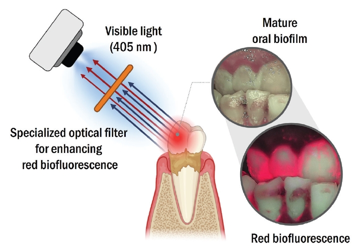
Schematic principle of oral biofilm detection using quantitative light-induced fluorescence (QLF).
However, studies applying biofluorescence technology to oral hygiene care (OHC) in hospitalized patients remain limited. Therefore, this study proposes a biofluorescence-guided oral care program termed Qray Monitoring Oral Care (QMOC), a systematic OHC program integrating real-time visualization, biofilm control, and reassessment. QMOC enables visualization of pathogenic biofilm and provides immediate visual feedback during intervention. This report describes the clinical application of QMOC for detecting and controlling pathogenic oral biofilm in a hospitalized geriatric patient with pneumonia and explores the potential role of biofluorescence for improving OHC in ward-based clinical settings.
Case Report
This report was approved by the Institutional Review Board (IRB) of Yonsei University Dental Hospital (IRB No. 2-2021-0053). Informed consent for publication was obtained from the patient’s legal representative due to the patient’s vulnerable condition.
A 78-year-old female hospitalized with pneumonia had a medical history of Parkinson’s disease and was undergoing hemodialysis for end-stage renal disease. She was bedridden due to severe physical decline and unable to communicate because of markedly impaired cognitive function. She also had dysphagia and received medications and enteral nutrition through a nasogastric tube.
A dentist visited the ward to perform an oral examination and establish an oral care plan. The Qraycam Pro (AIOBIO, Seoul, Korea) was used at the bedside to detect and monitor oral biofilm as part of QMOC. The oral cavity was extremely dry with noticeable oral malodor, reflecting poor oral hygiene. Thick oral biofilm was present on the teeth and at multiple oral sites, including the tongue dorsum, palate, and oral vestibule (Fig. 2). On visual inspection, the distribution and extent of mucosal biofilm were difficult to determine (Fig. 2 upper), whereas biofluorescence imaging (BFI) clearly revealed mucosal biofilm as RF, indicating pathogenic biofilm (Fig. 2 lower).
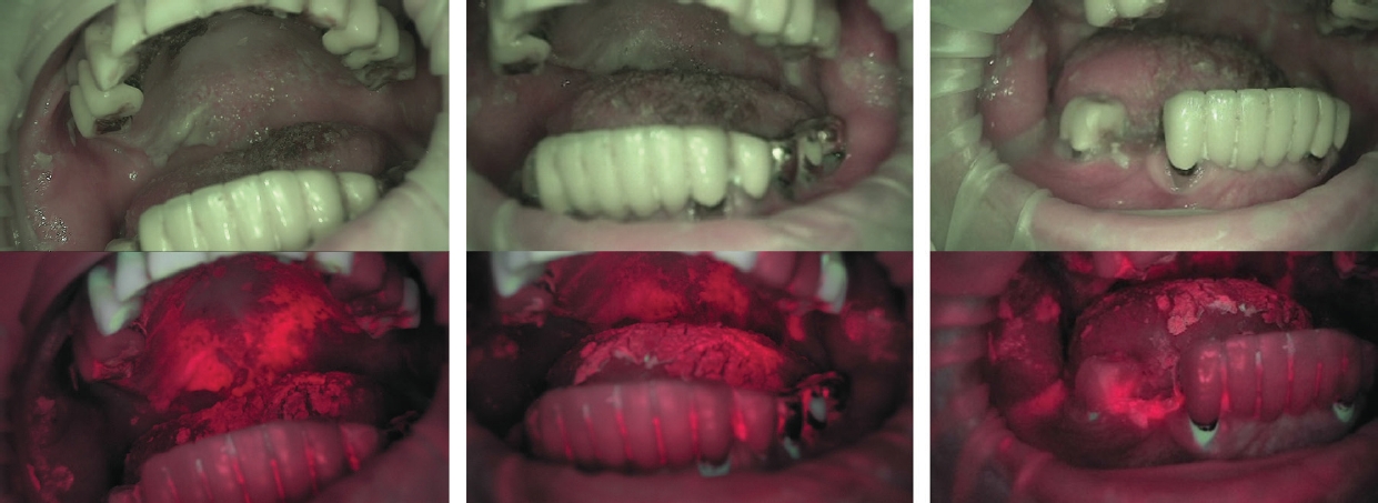
Extensive adherent oral biofilm visualized using quantitative light-induced fluorescence (QLF). Upper: white-light images; lower: biofluorescence images. Biofluorescence imaging enabled clearer identification of the distribution and extent of red biofluorescence-emitting biofilm.
OHC was performed by a trained dental hygienist according to a previously reported protocol [10]. The procedure focused on oral moisturizing and mechanical and chemical biofilm control. Artificial saliva substitute (Dry-mund, Dong-A Pharm, Seoul, Korea) and 0.12% chlorhexidine solution (Hexamedine, Bukwang Pharm, Seoul, Korea) were used as moisturizing and antimicrobial agents, respectively. Mechanical biofilm control was performed using a sponge brush (OK ORAL®, SOMOS International, Seoul, Korea), a manual toothbrush (Ruscello P20S, GC Fuji, Tokyo, Japan), and an interdental brush (Ruscello, GC Fuji, Tokyo, Japan).
Before oral care, the patient’s head was elevated to reduce the risk of aspiration. Because the oral cavity was severely dry, a moisturizing agent was first applied using a sponge brush. The sponge brush was then soaked in 0.12% chlorhexidine solution, excess fluid was removed with gauze, and the oral cavity was gently wiped to remove gross debris and contaminated secretions. Detached biofilm and debris were continuously suctioned using the bedside wall suction system. Tooth brushing was subsequently performed along the gingival margin and interdental areas. Finally, the oral cavity was wiped again with a sponge brush to collect residual contaminants, as oral rinsing was not feasible. During the procedure, QLF examination was used to detect residual RF biofilm in real time and guide OHC (Fig. 3). Although biofilm removal appeared complete on visual inspection, BFI revealed residual biofilm (Figs. 3B and C). Oral care was repeated until no biofilm was detected by either visual inspection or BFI (Fig. 3D). QMOC was continued until the targeted removable biofilm on both tooth surfaces and mucosal surfaces was no longer clinically detectable, as assessed by visual inspection and BFI. A moisturizing agent was subsequently applied to the oral tissues (Fig. 3D).
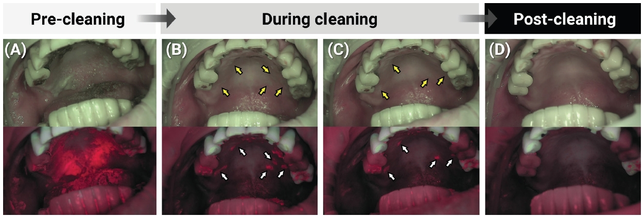
Clinical application of Qray Monitoring Oral Care (QMOC). A. Pre-cleaning. B and C. During cleaning. D. Post-cleaning. Upper panels: white-light images; lower panels: corresponding biofluorescence images. Residual adherent biofilm that was not clearly distinguishable under white light (yellow arrowheads) showed red fluorescence emission on biofluorescence imaging (white arrowheads).
To quantitatively assess the changes before and after QMOC, Multi-Chromatic Analysis (MCA) was additionally performed using LINKDENS (AIOBIO, Seoul, Korea). Figure 4 presents a representative application of MCA to adherent palatal biofilm. The analysis was conducted in nine oral regions, including the six dental sextants, right and left buccal mucosa, and palate. For each region, the pixel proportions of green, yellow, and red areas were calculated from the biofluorescence images. Values are presented as the mean ± standard deviation across all analyzed regions. The red area, classified in the MCA as a high-risk area indicating a relatively greater need for oral care, decreased from 17.29 ± 14.75% before QMOC to 1.23 ± 1.23% after QMOC, corresponding to a 16.06 percentage-point decrease and an approximately 92.9% relative reduction. Conversely, the green area, classified as a low-risk area, increased from 49.66 ± 26.95% before QMOC to 94.08 ± 4.71% after QMOC.
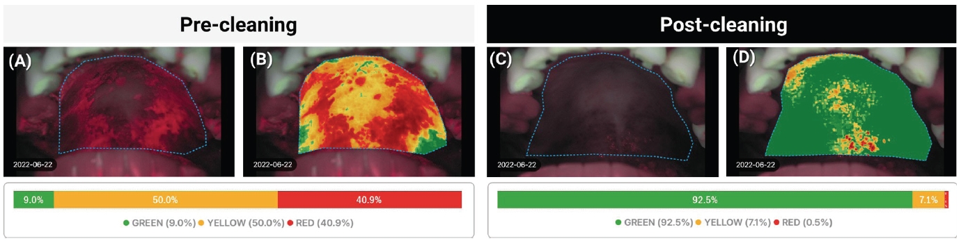
Representative application of Multi-Chromatic Analysis (MCA) for quantitative assessment of oral biofilm on the palate pre- and post- cleaning. A and C. Biofluorescence images with selected regions of interest (ROIs). B and D. Color-classified mapping images. The lower bar graphs show the pixel proportions of green, yellow, and red areas within each ROI.
Discussion
This report describes a clinical case demonstrating real-time detection and control of oral biofilm using biofluorescence in a hospitalized geriatric patient with pneumonia. QMOC enabled identification of mucosal biofilm that could be overlooked during visual inspection and allowed immediate confirmation of intervention outcomes. Thus, unlike conventional OHC, QMOC integrates real-time visualization, targeted biofilm removal, and objective post-intervention assessment within a systematic care process.
The QMOC protocol was based on QLF technology, which utilizes biofluorescence phenomena. Previous studies have demonstrated significant correlations between QLF-based plaque detection and conventional plaque-disclosing methods, particularly for mature and pathogenic dental biofilm [8,11-13]. Biofluorescencebased assessment can provide information not only on plaque distribution but also on its thickness and maturity through RF intensity, enabling more objective and reproducible assessment [8]. These findings suggest that RF not only serves as a useful visual indicator for detecting pathogenic biofilm but can also be used to evaluate the distribution and severity of biofilm through quantitative RF parameters, such as ΔR [14]. Moreover, RF has been observed on both dental hard tissues and soft tissues, including the tongue dorsum and oral mucosa, supporting its potential application beyond conventional dental plaque assessment [10,15-17].
However, most previous studies have involved relatively healthy adults. Highly dependent bedridden patients have distinct oral conditions, including reduced salivary flow, retained secretions, impaired self-cleansing, and frequent biofilm accumulation on mucosal surfaces [3,18,19]. Therefore, the applicability of biofluorescence-based biofilm detection should be further validated in this population. In the present case, RF functioned as a clinical decision-making indicator by identifying biofilm before care, guiding targeted removal, and allowing immediate reassessment after care. The clinical significance of this case therefore lies in translating a previously validated biofluorescence-based assessment approach into a realtime bedside OHC protocol for a vulnerable hospitalized patient.
Variability in OHC quality may arise from differences in operator expertise. Dental professionals are generally more experienced in assessing the oral environment and identifying biofilm [20], and previous studies have reported better oral or respiratory outcomes when professional oral care was delivered by dental personnel than when routine care was provided by general nursing staff [6,21]. These findings suggest that improved clinical outcomes may depend on the accurate identification and removal of pathogenic biofilm, as inadequate identification of biofilm targets may limit the effectiveness of oral care interventions.
Previous research has also emphasized the importance of objective oral assessment and high-quality OHC in reducing pulmonary infection and mortality in critically ill patients [22]. From this perspective, QMOC may help standardize the care process by providing a common visual criterion for identifying biofilm and confirming its removal. Real-time visualization may also facilitate detection of residual biofilm, including in anatomically difficult regions such as the oropharyngeal area, while reducing unnecessary repeated manipulation.
Meanwhile, mucosal biofilm appeared as a membranous substance adherent to the mucosa and was difficult to remove by simple cleansing. Such biofilm is commonly observed in long-term hospitalized patients with impaired oral function [18] and may serve as a reservoir for respiratory pathogens associated with pneumonia. Studies of respiratory specimens from patients with pneumonia have identified not only major respiratory pathogens but also periodontal anaerobic bacteria, including Prevotella and Fusobacterium [5], while oral Streptococcus species have been reported at higher abundance in bronchoalveolar lavage fluid from patients with pneumonia, particularly in suspected aspiration pneumonia [23]. These findings support the clinical importance of recognizing and managing mucosal biofilm in vulnerable patients.
RF in biofilm is primarily attributed to porphyrin metabolites produced by late-colonizing bacteria during the heme biosynthesis pathway in mature biofilm [9]. Previous studies have reported a greater abundance of Gramnegative anaerobic bacteria in RF-positive biofilms and increasing RF intensity with biofilm maturation [24]. Therefore, the RF observed in this case may reflect mature biofilm containing potentially pathogenic anaerobic bacteria, although the microbiological composition of the biofilm was not directly evaluated. Certain respiratory pathogens also exhibit intrinsic autofluorescence, suggesting additional potential for biofluorescence-based monitoring [25]. Therefore, BFI may represent a clinically relevant strategy for monitoring oral health in vulnerable patients. The QLF system is also relatively simple to operate, allowing healthcare providers and caregivers with limited dental expertise to recognize biofilm and confirm its removal.
This approach may be particularly relevant for patients at high risk of infection, including those who are critically ill, immunocompromised, or receiving radiation therapy. In such patients, poor oral hygiene may increase the microbial burden around medical devices such as endotracheal or nasogastric tubes and potentially contribute to systemic or respiratory infection [26]. By incorporating visual guidance and objective reassessment into OHC, QMOC may provide a more systematic approach to biofilm control in these vulnerable populations.
This study has several limitations. First, the microbiological characteristics of RF biofilm were not directly analyzed. Future studies using molecular approaches are needed to characterize the microbial composition in frail hospitalized patients. Second, this report was based on short-term observation in a single case; therefore, the long-term clinical impact of QMOC, including systemic infection, socioeconomic burden, and quality of life, could not be evaluated. Further studies are needed to investigate these aspects in broader patient populations and clinical settings.
For broader clinical implementation of QMOC, both device accessibility and user education should be considered. To enable non-dental healthcare professionals and caregivers to consistently use biofluorescence images in oral care, basic training in image interpretation is needed. Such training may also facilitate multidisciplinary communication by providing a common visual standard for recognizing and sharing oral hygiene status. Although the initial cost of QLF devices may be a practical barrier, this burden may be reduced by using more affordable screening devices, such as Qscan Plus™ (AIOBIO, Seoul, Korea), in combination with smartphone-based imaging to record and share biofluorescence images.
In conclusion, biofluorescence-guided oral care may enable real-time detection of pathogenic oral biofilm, including residual biofilm overlooked by visual inspection, and objective evaluation of intervention outcomes. These findings support the potential role of biofluorescence in improving OHC in clinical practice.
Notes
Conflicts of Interest
None