Efficacy of preoperative sclerotherapy in the surgical management of buccal venous hemangioma: A case report
Article information

Abstract
Sclerotherapy has emerged as a valuable adjunctive therapy in the management of venous hemangioma. Sclerosant agents induce endothelial damage, leading to fibrosis and obliteration of the vascular lumen, ultimately reducing blood flow to the lesion. In this case report, we present a patient with a venous hemangioma of the buccal mucosa who underwent four sessions of sclerotherapy with limited lesion regression. However, during subsequent surgical resection, intraoperative bleeding was minimal and postoperative healing was uneventful, suggesting that sclerotherapy contributed significantly to surgical hemostasis. This report highlights the potential role of preoperative sclerotherapy in facilitating the safe resection of oral venous hemangiomas.
Introduction
Oral venous hemangiomas are benign tumors that develop due to endothelial cell proliferation. Although oral venous hemangiomas are relatively rare, when they do occur, they show a predilection for the lips, tongue, buccal mucosa, and palate. According to Mulliken and Glowacki classification system in 1982 [1], “hemangiomas” are true neoplasms characterized by proliferation and increased rates of endothelial cell turnover, while “vascular malformations” are localized anomalies due to defects in vascular morphogenesis with normal rates of cell turnover.
Therapeutic strategies for oral venous hemangiomas include pharmacologic agents such as beta-blockers (e.g., propranolol), corticosteroids, surgical excision, and more recently, sclerotherapy using agents such as sodium tetradecyl sulfate (STS). Sclerosing agents work by damaging the vascular endothelium, causing thrombosis and subsequent fibrosis, leading to vascular occlusion. In 2018, systematic review by Kristina R. Asdahl et al. reported that sclerotherapy is a safe, efficient, and good treatment method for venous malformation. However, complications including swelling, pain, inflammation, and skin blistering can occur [2]. Notably, the incidence of facial nerve paralysis is higher with ethanol (up to 18%) compared to other sclerosants (0–6%). Additional studies support its efficacy across a range of vascular anomalies, particularly when used in conjunction with surgical intervention [2,3].
This report evaluates the clinical impact of STS sclerotherapy as a preoperative intervention for a buccal ve-nous hemangioma, with specific focus on its effects on lesion vascularity and intraoperative hemostasis.
Case Report
A 57-year-old male was referred to the department with a persistent submucosal mass on the left buccal mucosa (Fig. 1). Fine-needle aspiration yielded bloody fluid, consistent with a vascular lesion. The lesion was diagnosed clinically as an oral venous hemangioma.
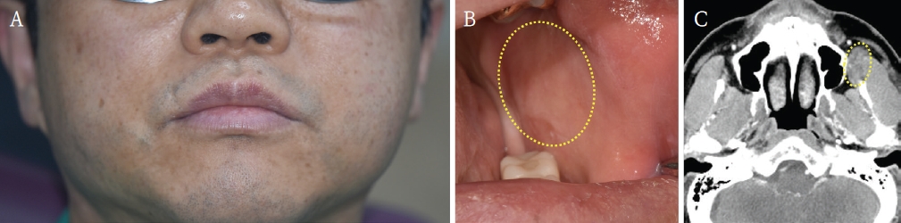
A and B. Clinical images of extra/intraoral lesion site. C. Enhanced facial computed tomography imaging shows hypoattenuation lesion adjacent to the left buccal mucosa.
Sclerotherapy was initiated using 3% sodium tetradecyl sulfate (Fibrovein 61.22 mg/mL; 30 mg as STS, STD Pharmaceutical Product Ltd., Hereford, UK). 1cc of 3% Fibrovein was used on the lesion for 4 months at 1 month intervals. Reduction in lesion size was noted after the first two sessions, but subsequent injections yielded no further significant change.
Given the plateau in response, surgical excision was planned under local anesthesia. Routine draping was performed after injection of 1:100,000 lidocaine mixed with epinephrine. To avoid injury to Stensen’s duct, a horizontal mucosal incision (1.5 cm) was made horizontally on the buccal mucosa below the duct (Fig. 2A). Blunt dissection was used to approach the buccal space, and buccal fat was pulled to expose the lesion to prevent bleeding (Fig. 2B). The lesion was resected through blunt dissection, and the inside of the mass appeared to be adhered to buccal fat (Figs. 2C and D). The lesion was carefully excised and bleeding during surgery was not notable (Fig. 2E). Postoperative healing was uneventful.

A. Incision was made horizontally below the Stensen’s duct. B. Buccal fat was exposed through blunt dissection. C and D. The excised lesion measured approximately 3.5 cm in size and demonstrated adhesion to the buccal fat. E. Postoperative image shows no remarkable findings.
Gross examination revealed pearl-like, firm nodules on both the external and internal surfaces of the lesion, measuring 2 mm and 5 mm in diameter, respectively (Fig. 3).
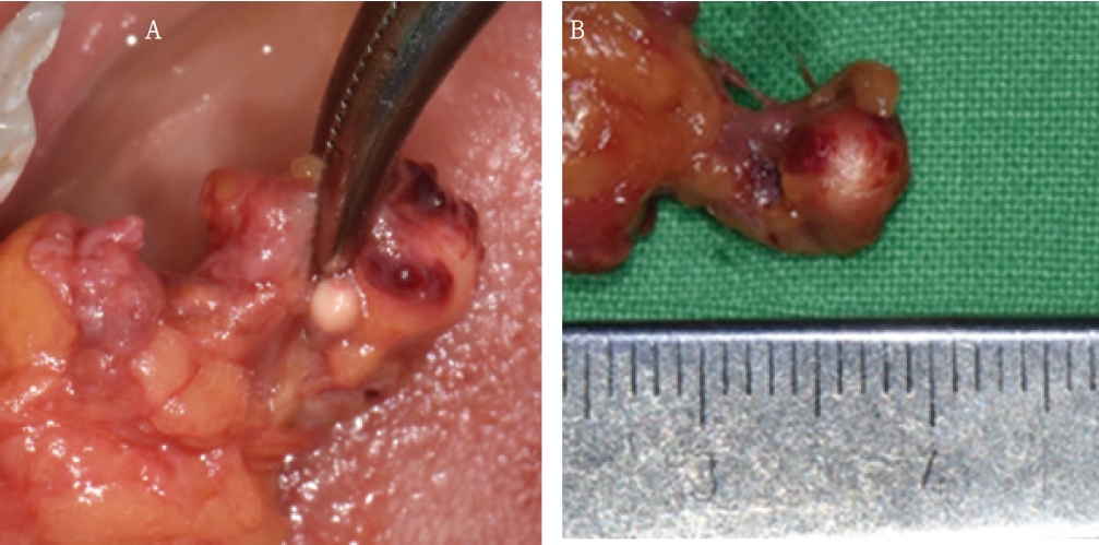
A. Pearl-like, firm nodules on the lesion. B. Each nodule measured 2 mm and 5 mm, respectively.
Histopathological analysis confirmed the diagnosis of oral venous hemangioma. Microscopy revealed fibrotic and partially calcified venous channels surrounded by congested capillaries, with the pearl-like nodules identified as dystrophic calcifications within sclerosed vascular lumens (Fig. 4).
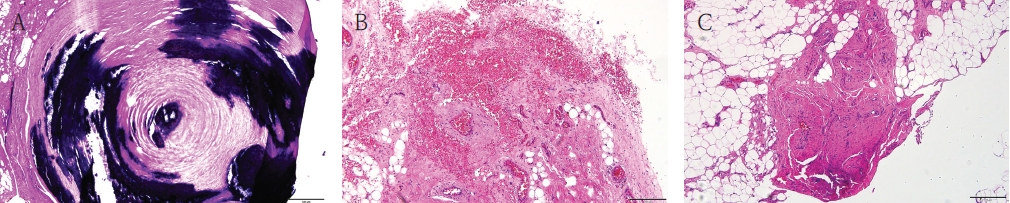
Histopathological findings of the resected oral venous hemangioma following STS sclerotherapy. A. Blood vessel with dystrophic calcification (H&E stain; original magnification x100). B. Venous hemangioma characterized by dilated vascular spaces lined by flattened endothelial cells (H&E stain; original magnification x200). C. Vascularized fibroadipose tissue containing small-caliber vascular channels (H&E stain; original magnification x200).
Discussion
Sclerotherapy using sodium tetradecyl sulfate pro-motes endothelial disruption, thrombotic occlusion, and subsequent fibrosis of low-flow vascular anomalies. In the current case, while complete lesion regression was not achieved through sclerotherapy alone, the pretreatment effectively diminished vascularity and provided a near bloodless operative field, which is a considerable advantage in highly vascularized intraoral sites.
Numerous studies support the utility of sclerotherapy as a minimally invasive modality in managing vascular lesions of the head and neck. Resmije et al. documented favorable outcomes with minimal complications in oral hemangiomas treated with sclerosants [4]. However, complete lesion resolution remains challenging in venous malformations due to the complexity of their vascular channels [4,5].
Various sclerosants have been employed for the treatment of oral venous hemangiomas, each exhibiting distinct mechanisms of action, efficacy profiles, and complication risks. STS, a detergent-based sclerosant, has been widely used in head and neck lesions due to its relatively favorable safety profile and moderate efficacy [6]. Polidocanol, another detergent agent, has demonstrated lower post-procedural discomfort and inflammation, making it particularly suitable for lesions in sensitive anatomical zones [7]. STS and polidocanol had lower success rates (35–50%) but were associated with fewer adverse events [8]. In contrast, absolute ethanol, a potent dehydrating agent, has shown superior efficacy in lesion regression (up to 66% complete response) but carries a significantly higher risk profile, including dermal necrosis, neuropathy, and systemic toxicity [2].
STS offers several advantages in the treatment of oral venous hemangioma. Compared with ethanol, STS causes less tissue necrosis, nerve injury, and systemic toxicity, making it particularly suitable for lesions located in anatomically sensitive regions of the oral cavity [9]. Common adverse effects include transient pain, swelling, localized inflammation, and occasional superficial ulceration, which are generally self-limiting [9,11]. In contrast, ethanol-based sclerotherapy has been associated with significantly higher rates of severe complications including skin necrosis and cranial nerve injury [2,3]. Therefore, STS is often preferred when preservation of adjacent functional structures is critical. Previous studies have demonstrated the safe use of STS in various intraoral locations including the tongue, lips, buccal mucosa, and floor of the mouth [10]. Although temporary swelling following injection may be more clinically significant in the tongue because of the potential risk of airway compromise, STS has generally shown a favorable safety profile when administered in appropriate doses with careful postoperative monitoring [10,11]. Therefore, STS can be considered a reliable sclerosant for low-flow venous hemangiomas involving major oral soft tissues [9,10].
A systematic review and meta-analysis by De Maria et al. [12] further highlighted that the overall treatment success rate across sclerosants is approximately 83%, with ethanol outperforming other agents in efficacy but at the cost of increased adverse event incidence (up to 18%). Meanwhile, STS and polidocanol reported lower regression rates (35–50%) but were associated with fewer complications, making them preferable in functionally or aesthetically critical areas. Bleomycin, a cytotoxic antineoplastic agent, has been employed primarily in pediatric populations with promising outcomes, though concerns regarding cumulative pulmonary toxicity remain [3]. Recent innovations such as bleomycin electrosclerotherapy (BEST) have demonstrated enhanced lesion control via electroporation-facilitated drug delivery, showing potential in treatment-resistant cases [13].
In the present case, histological evaluation revealed fibrotic transformation and dystrophic calcification of vascular structures, consistent with the tissue response to repetitive sclerotherapy. These findings support the concept that even when size reduction is suboptimal, pretreatment with sclerosants can significantly mitigate intraoperative bleeding and facilitate safe resection. Ultimately, the selection of an optimal sclerosant should be individualized, taking into account anatomical location, lesion size and flow characteristics, as well as patientspecific factors such as age, comorbidities, and tolerance to potential side effects. For intraoral venous hemangioma requiring surgical intervention, STS remains a clinically effective and well-tolerated option, particularly when used in a multimodal treatment strategy.
At our institution, treatment selection for oral venous hemangioma is based on lesion size, anatomical location, symptoms, and expected surgical morbidity. Small, well-circumscribed lesions (<1 cm) are generally managed by surgical excision alone when complete removal can be achieved with minimal functional or esthetic compromise. Diffuse lesions or lesions involving critical structures are initially treated with sclerotherapy. For medium-sized lesions (approximately 1–3 cm) in which surgical excision is anticipated to be associated with increased bleeding risk, preoperative sclerotherapy is performed to reduce lesion vascularity, followed by delayed surgical excision if residual disease persists. This combined approach aims to maximize lesion control while minimizing operative morbidity [9,11].
While sclerotherapy alone may not achieve complete lesion regression in all oral venous hemangiomas, its preoperative application can substantially reduce intraoperative bleeding and enhance surgical feasibility. This case supports the incorporation of sodium tetradecyl sulfate sclerotherapy into a comprehensive management approach for intraoral vascular lesions.
Notes
Conflicts of Interest
None