콘빔CT에서 우연히 발견된 상악동 및 안면 피하 연조직 이물질 5례
Incidental foreign bodies beyond the jaws on cone-beam computed tomography: Report of 5 cases
Article information

Trans Abstract
Cone-beam computed tomography is used to evaluate the teeth and jaws and for orthodontic assessment and implant treatment planning. Compared with panoramic radiography, it provides cross-sectional imaging and a large field of view, enabling superimposition-free assessment beyond the dentoalveolar region. Consequently, incidental findings unrelated to the original indication are commonly encountered and have been reported in up to 94% of examinations. These findings include anatomic variants, soft-tissue calcifications, foreign bodies, and lesions that may warrant clinical management, often in sinonasal spaces and other soft tissues. This report showed 5 cases of incidentally detected foreign bodies. Three foreign bodies in the maxillary sinus or facial soft tissue were not recognized on panoramic radiographs and were detected on cone-beam computed tomography. Two suspected on panoramic radiography were further characterized with definitive localization and morphology. These cases emphasized that careful review of the entire image volume was essential to prevent missed foreign bodies.
서론
콘빔CT는 치아 및 악골부위 골병소진단에 유용한 검사이다. 파노라마방사선검사에서 제공되지 않는 3차원 정보를 제공하며, 관심영역(field-of-view, FOV)의 크기에 따라 악골 외에 비강, 상악동, 악관절, 경부등 두경부영역을 광범위하게 관찰할 수 있다. 이러한 장점으로 치과영역에서 콘빔CT는 필수적인 영상검사가 되었다.
촬영된 콘빔CT영상에서 원래 목적과는 관련없는 소견들이 우연히 발견되기도 한다[1-4]. 우연히 관찰된 소견(incidental finding)이 얼마나 흔하게 발견되는지는 보고서마다 다양하지만, 최대 94%에서 발견되며, 콘빔CT의 하나의 단면상에서 평균 1-3개의 소견이 동시에 발견되기도 한다[1,2]. 우연히 관찰된 소견의 양상은 해부학적 변이부터 익숙하지 않은 이물질, 임상적으로 주요한 병변까지 다양하다. 특히, 치료나 추적검사가 필요한 임상적으로 의미있는 소견(clinically significant incidental finding)인 경우에는 보다 주의 깊은 영상 판독이 이루어져야 하며, 적절한 치료계획을 세울 수 있어야 한다.
이에 본 저자는 콘빔CT영상에서 우연히 발견되었지만, 특별한 치료를 요하지 않는 이물질 증례와 외과적 치료가 필요한 임상적으로 의미있는 이물질 증례를 보고하고자 한다. 이를 통하여 치아 및 악골외 부위에서 관찰될 수 있는 이물질 소견들을 공유하고, 이들 소견에 대한 판독과 콘빔CT검사의 임상적 유효성을 강조하고 한다.
증례
증례 1
20대 여자환자가 윗 입술의 불편감을 주소로 내원하였다. 파노라마방사선영상에서 관련된 이상소견이나 병변이 관찰되지 않았다(Fig. 1A). 과거병력 청취에서 교통사고로 인한 윗 입술 열상의 봉합 병력이 있었음을 파악한 후, 외상 후 연조직 섬유화로 진단되었다. 1년 후, 이물감을 호소하면서 환자는 재내원하였다. 파노라마방사선영상에서는 여전히 이상소견이 관찰되지 않았기에, 콘빔CT검사를 시행하였다. 윗입술과 상악 전치사이 연조직부위에서 블럭모양의 방사선불투과성 이물질이 발견되었다(Fig. 1B). 이물질 제거를 위한 외과적 수술이 시행되었고, 술 후 해당 이물질은 유리조각임이 확인되었다.
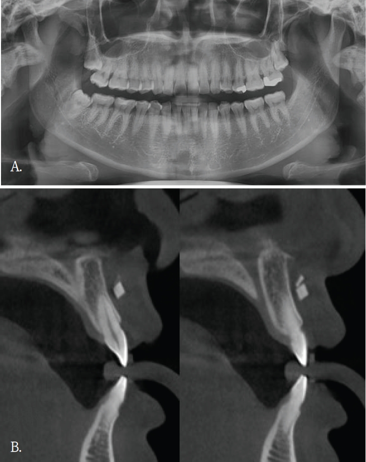
A. Panoramic radiograph shows no definite abnormality in the maxillary anterior region. B. Cross-sectional cone-beam computed tomographic image of the maxillary anterior alveolar region demonstrates a small block-shaped radiopaque foreign body in the labial soft tissue, without intraosseous involvement.
증례 2
50대 여자환자가 상악 전치부 임플란트치료를 위해서 콘빔 CT검사를 시행하였다. 파노라마방사선영상에서는 해당 부위에 특이 소견이 뚜렷하지 않았다(Fig. 2A). 그러나 콘빔CT의 축상면 및 횡단면 영상에서 양측 상악동 전벽에 인접한 안면 피하 연조직을 따라, 반달모양의 경계가 명확하고 내부가 균질한 음영을 보이는 구조물이 관찰되었다(Figs. 2B and C). 해당 소견은 주된 촬영 목적과 무관하였고 양측에서 대칭적으로 관찰되었으며 관련 증상이 동반되지 않았다. 임상 소견 및 영상 소견을 종합할 때 병변 가능성은 낮다고 판단하였고, 위치와 형태를 고려하여 피하에 주입된 미용필러에 의한 소견으로 판단하였다. 이에 추가 처치는 시행하지 않았다.
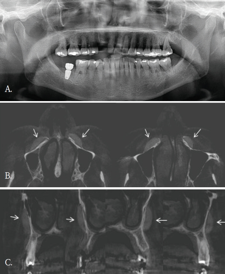
A. Panoramic radiograph shows no definite abnormality in the maxilla. B. Axial cone-beam computed tomographic (CBCT) image shows crescent-shaped radiopaque foreign bodies along the anterior walls of the bilateral maxillary sinuses (arrows). C. Cross-sectional CBCT image also demonstrates the same findings on a different plane (arrows).
증례 3
좌측 안면 부종으로 타원에서 의뢰된 30대 여자환자의 파노라마방사선영상에서 좌측 하악체 및 하연부를 따라 길이가 약 7cm로 측정된, 가는 선 모양의 방사선불투과성 이물질이 관찰되었다(Fig. 3A). 그러나 파노라마방사선영상에서 이물질의 협·설측 위치 및 하악골 내 포함여부를 명확히 판단하기 어려웠다. 이에 위치 및 주변 구조물과의 관계를 평가하기 위해 콘빔CT검사를 시행하였다. 콘빔CT에서 이물질은 좌측 하악체 외측 피질골 바깥의 협측 연조직에서 관찰되었다(Fig. 3B). 영상소견을 바탕으로 구강 내 접근을 통해 외과적 제거술이 시행 되었고 제거된 이물질은 부러진 주사바늘로 확인되었다.
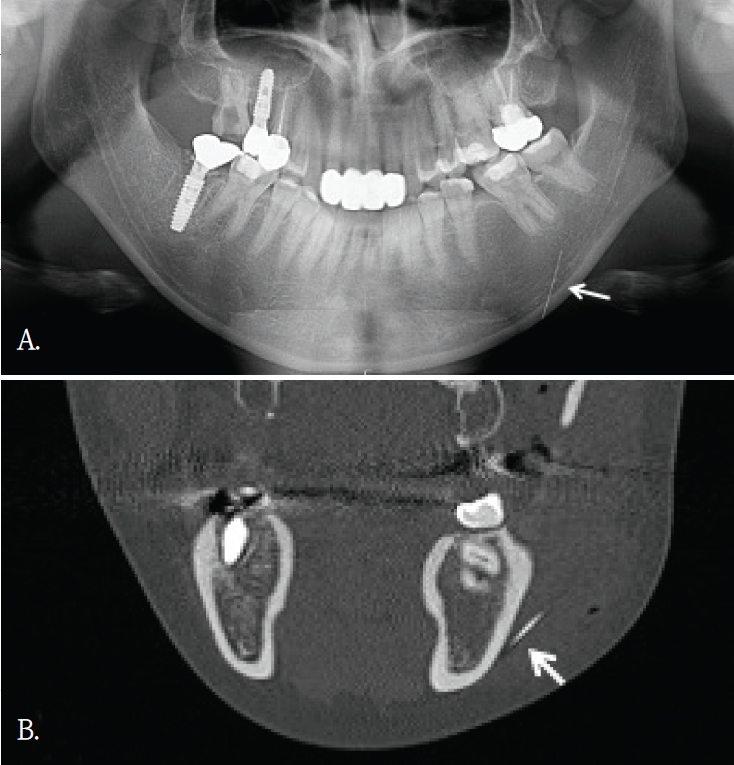
A. Panoramic radiograph shows a long, thin, linear radiopaque foreign body along the left mandibular body and inferior border (arrow). B. Coronal cone-beam computed tomographic image demonstrates that the linear radiopaque foreign body is located in the buccal soft tissue lateral to the mandible (arrow).
증례 4
임플란트 식립 중 식립체가 시야에서 소실되어 위치 확인을 위해 60대 여자환자에서 파노라마방사선검사를 시행하였다. 파노라마방사선영상에서 임플란트 식립체로 추정되는 금속성 방사선불투과성 구조물이 우측 비강 부위에 중첩되어 관찰되었고 영상에서 비강 하연 부근에 위치한 것처럼 보였다(Fig. 4A). 이에 비강 및 상악 치조골과의 정확한 위치 관계를 평가하기 위해 콘빔CT 검사를 시행하였다. 콘빔CT에서 해당 구조물은 고음영 금속성 구조물로 확인되었으며, 상악 우측 전치부 측와 부위의 순측 연조직에서 관찰되었다(Figs. 4B and C). 또한 비강저 및 상악 치조골 피질골의 연속성이 보존되어 구조 발치와가 관찰되었다(Fig. 5A). 이러한 소견은 좌측 상악동 점막의 염증성 변화 가능성을 시사하였다. 상악동 병변의 범위 및 발치와와 상악동하연의 관계를 평가하기 위해 콘빔CT검사를 시행하였다. 콘빔CT에서 좌측 상악동 내에 연조직음영에 의한 혼탁이 관찰되었으며, 이는 상악동의 약2/3이상을 채우는 양상이었고 상악동 소공주변까지 연장되어 보였다. 또한, 상악동 전하방에서 경계가 명확한 작은 타원형의 균질한 고음영 구조물이 관찰되었으며, 골밀도에 해당하는 방사선불투과성 이물질로 판단되었다(Figs. 5B and C). 해당 고음영 소견은 파노라마방사선영상에서는 명확히 구분되지 않았다. 환자의 발치 병력을 고려할 때 해당 구조물은 상악동 내로 이동한 잔존치근이 의심되었으며, 외과적 제거를 시행하였다. 제거된 이물질은 상악동 내 잔존 치근으로 확인되었다.
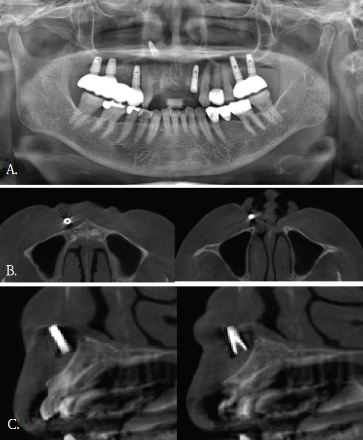
A. Panoramic radiograph shows an implant-shaped foreign body superior to the maxillary anterior region and appearing superimposed on the nasal cavity. B. Axial cone-beam computed tomographic (CBCT) image demonstrates that the foreign body is located in the labial soft tissue, without involvement of the nasal cavity. C. Cross-sectional CBCT image demonstrates that the foreign body is located in the labial soft tissue rather than in the maxillary anterior alveolar bone.
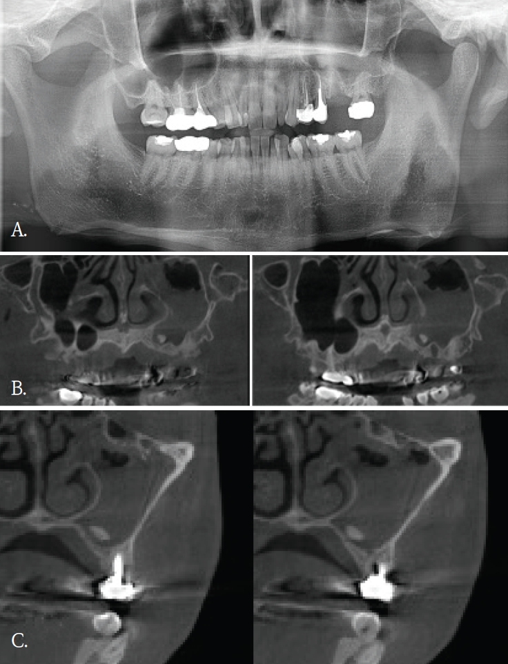
A. Panoramic radiograph. An extraction socket of the left maxillary first molar is noted. Diffuse haziness of the left maxillary sinus is observed; no other definite abnormality is identified. B. Panoramic reconstruction, C. cross-sectional cone-beam computed tomographic images. Soft-tissue density nearly fills the left maxillary sinus. A small radiopaque foreign body is identified at the floor of the sinus.
고찰
콘빔CT는 해부학적 구조물이나 이물질 위치를 확인하고, 인접 구조물과의 관계를 파악하는데 유리하다. 2차원 영상에서 발생되는 중첩을 줄이고, 3차원적인 위치정보를 제공함으로써 치과 임플란트 치료계획이나 치아 교정 진단과정에서 필수적인 영상검사이다. 또한, 임플란트 식립이나 매복치 발거등 외과적 치료과정에서도 진입 경로 결정에 도움을 주어 술 후 합병증을 감소시킨다.
콘빔CT의 원래 촬영 목적과는 관계없이, 우연히 발견되는 소견들이 있다. 이러한 소견의 빈도는 24.6~94.3%로 보고마다 매우 다양하다[1,2]. 소아 및 청소년연령군에서의 빈도도 0.4~80.3%로 다양하지만 매우 자주 발견된다[3]. 발견되는 부위는 치아 및 치조골을 벗어난 부위가 대부분으로 주로 부비동 및 비강, 혈관, 연조직, 경추이다[1-3]. 그러므로, 콘빔CT 검사할 때 촬영목적 부위 뿐 만 아니라 그 외 다른 부위의 판독도 충분히 이루어져야 한다[2,4,8]. 특히 어린 환자에서는 방사선방어를 고려하여, 관심부위 뿐 만 아니라 콘빔CT영상에 포함된 모든 부위에서 영상판독이 충분히 이루어져야 한다[3]. 우연히 발견되는 소견의 양상도 매우 다양하다. 부비동 및 비강에서는 점막비후, 점액저류낭, 비중격만곡이 많이 발견되고, 혈관부위에서는 경동맥석회화가 주로 발견된다. 연조직에서는 편도석, 경동설골인대골화가 주로 발견되는 소견이다[1-3]. 대부분 해부학적 변이이거나 의학적 조치가 필요하지 않은 경우이다.
하지만 추적검사나 치료가 필요한 임상적으로 의미가 있는 소견들도 매우 다양한 빈도로 발견된다. 최소 0.4%에서 최대 37%까지로 전체의 약 1/6-1/3 정도에서 발견되어 주의깊은 콘빔CT 영상판독이 필요하다. 이들 소견중에는 치근단병소, 치성낭, 종양도 있었으며[1-3], 오랜 기간 동안 인지하지 못했던 이물질도 발견된다[1,4,6,8].
우연히 발견된 이물질들은 치조골 및 악골 부위가 아닌 다른 부위에서 많이 관찰되었다. 교정목적으로 촬영된 콘빔CT에서 비강내에 플라스틱 이물질이 우연히 발견되어 이비인후과 의뢰 후 이물질을 제거한 증례보고가 있다[4]. 증상이 없어서 이물질의 유무를 알 수 없었지만, 콘빔CT에서 비강내 이물질이 확인되어 외과적으로 제거한 증례보고도 있다[5,6]. 증상이 있어 촬영한 상악 견치 치근단방사선사진에서 모호한 방사선불투과성 이물질이 관찰되어, 이후 촬영한 콘빔CT에서 이물질이 유리조각으로 확인된 증례보고도 있다[8]. 이 증례는 유리같은 이물질은 치근단이나 파노라마방사선사진에서는 명확하게 관찰되지 않았기에 콘빔CT검사가 매우 유용하였던 경우이다.
증례 1에서도 초진시 파노라마영상에서는 유리조각을 관찰할 수 없었기에 연조직 병변으로 진단되었으나, 재내원후 촬영된 콘빔CT영상에서 연조직내에 있었던 이물질임으로 확인된 경우이다. 윗입술에 있었던 유리조각이 방사선불투과성 물질이였지만, 상악 전치부와 중첩되어 파노라마방사선영상에서 뚜렷하게 구분되지 않았다.
증례 5와 같이 파노라마방사선영상에서 모호하게 관찰되었거나 증례 3과 4같이 파노라마방사선영상에서 협-설위치를 명확하게 알 수 없었던 경우, 콘빔CT영상에서 임상적으로 의미있는 이물질임을 확인되었고 외과적 치료가 시행되었던 증례들이다. 특히, 이들 소견은 악골이 아닌 안면 연조직 부위에서 관찰되어, 주의 깊은 영상판독의 중요성을 다시금 일깨워 준다.
한편, 안면미용시술을 받은 환자들이 치과에 많이 내원하면서 우연히 발견되는 이물질 소견 중 하나가 미용 필러이다[9]. 치과의사들에게 아직은 익숙하지 않은 방사선영상 소견이고 부위도 치아나 악골이 아닌 연조직부위에서 관찰되기에 영상 판독에서 배제될 가능성이 있다. 미용필러도 파노라마방사선 영상에서보다는 콘빔CT영상에서 위치나 형태, 양상을 잘 관찰할 수 있다.
본 증례들은 파노라마방사선영상에서 인지되지 않았던 이물질 3증례와 파노라마방사선영상에서 의심된 이물질이 콘빔 CT에서 위치와 형태가 명확히 규명된 2증례를 포함한다. 이를 통해 악안면 콘빔CT 판독 시 촬영 목적 부위에 국한하지 않고 영상에 포함된 전체 영역을 체계적으로 검토함으로써, 이물질과 같은 우연히 발견되는 소견이 누락되는 것을 줄일 수 있음을 알았다. 또한 추가 촬영이나 불필요한 검사로 인한 방사선 노출을 예방하여, 콘빔CT 검사의 정당성과 임상적 유효성을 강화하는 뒷받침이 됨을 알았다.
Notes
Conflicts of Interest
None