파노라마방사선영상에서 광범위하게 관찰된 두경부의 동맥석회화 증례
A case of extensive arterial calcification in the head and neck observed in panoramic radiography
Article information
Trans Abstract
Arterial calcification, which involves calcium deposits in the arterial walls, can be categorized into intimal and medial types. Intimal calcification is often linked to atherosclerotic plaques, while medial calcification is associated with aging and hypercalcemia. This case involves a 52-year-old male with severe chronic kidney disease, who is undergoing dialysis treatment. Panoramic radiography revealed arterial calcifications in common carotid, external carotid, internal carotid, lingual, facial, superior labial, and maxillary arteries, bilaterally. Such findings on a panoramic radiograph are rare but quite plausible in patients with severe chronic kidney disease. Panoramic radiographs may not always capture such calcifications, and CT or cone-beam CT might reveal more cases. When detected, the possibility of chronic kidney disease or other hypercalcemia-related conditions should be considered.
서론
동맥석회화(arterial calcification)는 동맥의 내벽에 칼슘이 침착되는 현상으로 두 가지 유형으로 나타난다. 하나는 동맥혈관의 내막이 석회화되는 것(intimal calcification)이고 다른 하나는 중막이 석회화되는 것(medial calcification)이다. 내막 석회화는 주로 죽상경화반(atherosclerotic plaque)과 관련이 높다[1,2]. 중막 석회화는 노화 및 고칼슘혈증(hypercalcemia)과 관련이 높으며 Mönckeberg's arteriosclerosis라고도 한다[3-6]. 죽상경화반에 의한 내막 석회화는 이형성석회화(dystrophic calcification)에 속하며 고칼슘혈증에 의한 동맥석회화는 전이석회화(metastatic calcification)의 일종이다.
동맥석회화는 악안면영역에서는 경동맥(carotid artery) 및 안면동맥(facial artery)에 흔히 발생한다. 죽상경화반에 의한 석회화는 경동맥의 분지부(bifurcation)에 주로 관찰되며 고칼슘혈증에 의한 석회화는 경동맥 및 안면동맥에 광범위하게 관찰되는 경우가 많다.
죽상경화반에 의한 경동맥의 석회화는 뇌졸중과 연관성이 높다. 파노라마방사선영상은 비록 CT영상이나 CBCT영상에 비하여 경동맥 석회화의 검출률은 낮지만[7] 촬영빈도가 매우 높기 때문에 뇌졸중 위험이 있는 환자를 감지하는 데 유용한 도구가 될 수 있다[8].
죽상경화반의 석회화는 혈관벽에 불규칙한 석회화를 보여 파노라마방사선영상에서도 불규칙하게 관찰되며[9] 고칼슘혈증에 의한 석회화는 파노라마방사선영상에서 기찻길 모양 또는 원형으로 관찰된다[6,10].
파노라마방사선영상을 이용하여 죽상경화반에 의해 발생한 경동맥의 석회화를 보고한 증례나 연구는 다수이다. 그러나 파노라마방사선영상에서 전이석회화의 일종인 고칼슘혈증에 의한 동맥석회화는 보고가 매우 드물어, 외경동맥 및 그 가지에 발생한 증례를 소개하고자 한다.
증례
52세 남자 환자가 남아있는 치아를 모두 발치하고 틀니를 하고 싶다는 주소로 내원하였다. 환자는 만성신부전 때문에 일주일에 3회 신장투석을 받는 신장 2급 장애인이었으며 고혈압으로 인해 약물 치료를 받고 있었다. 과거에 결핵이 완치된 병력이 있었다.
임상 검사 결과, 상악에는 우측 견치, 우측 제3대구치, 좌측 견치가, 하악에는 우측 견치, 우측 제1소구치, 좌측 견치가 잔존하였으며, 모두 치아우식증이 있었고 전반적으로 심한 치은 퇴축이 관찰되었다. 파노라마 방사선 영상에서는 전반적으로 심한 치조골 흡수가 관찰되었다. 상악 우측 제3대구치와 하악 좌측 견치는 부유치였으며, 하악 제1소구치에는 치근단 희박화 골염이 관찰되었다.
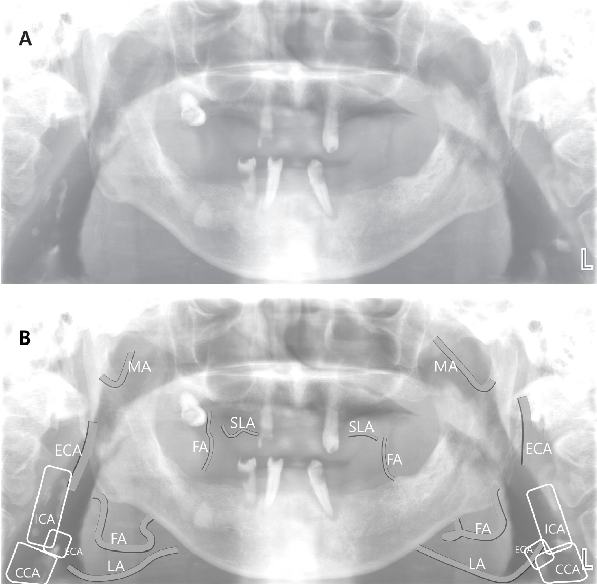
양측 경추 2번, 3번 및 4번의 전방에 다수의 석회화 물질들이 수직선상에 불규칙하게 배열된 것이 관찰되었다. 이 중 하방의 상대적으로 넓은 폭경 부분은 총경동맥(common carotid artery)의 석회화로 진단하였다. 양측 후두덮개 부위에 석회화 부분이 중첩되어 있었고 이 부분이 약간 전방으로 돌출되어 외경동맥(external carotid artery)의 시작부분으로 판단하였으며 그 후방 및 상방의 두꺼운 불규칙한 석회화부분은 내경동맥(internal carotid artery)의 석회화로 진단하였다. 이들은 불규칙한 석회화를 보여 죽상경화반에 의한 내막 석회화로 진단하였다(Figure 1).

A, Panoramic radiograph showing artery calcifcation at the bilateral carotid bifurcation areas, bilateral external carotid arteries, and their branches, including the lingual, facial, superior labial, and maxillary arteries. B, Drawing of the arteries seen in A (MA: maxillary artery; SLA: superior labial artery; FA: facial artery; LA: lingual artery; ECA: external carotid argery; ICA: Internal carotid artery; CCA: common carotid artery)
양측 구치부 하악체 하방, 협부, 상순, 하악지 후방 및 하악 절흔 부위의 연조직에도 한 쌍의 가느다란 방사선불투과성 선들이 기찻길 모양으로 다수 관찰되었다. 이 부위의 석회화는 아래로부터 설동맥(lingual artery), 안면동맥의 하방 부위, 안면동맥의 협측 부위, 안면동맥의 분지인 상순동맥(upper labial artery), 악동맥(maxillary artery)에 발생한 동맥석회화로 진단하였다. 그 밖에도 하악 하방에 다수의 가느다란 선들이 관찰되었는데 이것들은 설동맥 및 안면동맥의 가지들로 판단하였다. 이들은 모두 기찻길 모양의 석회화를 보여 중막 석회화로 진단하였다(Figure 1).
토의
일반적으로 이차성 부갑상선항진증에서는 혈 중 칼슘농도가 낮지만 이차성 부갑상선항진증이 오래 지속되다보면 부갑상선이 자율성을 가지고 과도한 부갑상선 호르몬을 분비하여 그 결과 고칼슘혈증을 나타나게 된다. 이것을 삼차성 부갑상선항진증이라고 한다[4,5,11]. 이 환자는 혈액투석을 받는 신장2급 장애인이있으며 신장투석을 받는 중증 신부전증환자가 여기에 속한다. 따라서 이 환자의 삼차선 부갑상선항진증에 의한 혈관석회화로 판단된다.
파노라마방사선영상에서 혈관석회화에 대한 연구는 주로 경동맥 석회화에 대한 것이 대부분이며[7-9] 한국인을 대상으로 한 논문도 대부분 경동맥 석회화에 대한 것이다[1,2,6]. Suarez-Cunqueiro 등[10]은 2002년 파노라마방사선영상에서 외경동맥 분지의 석회화를 발표하면서 최초의 증례보고라고 주장하였다. 한국인을 대상으로 한 논문은 PubMed와 한국학술지인용색인을 통해 검색해 보았을 때 단 하나의 연구도 발견할 수 없었다. 이번 증례는 파노라마방사선영상에서 한국인을 대상으로 석회화된 외경동맥의 분지를 설명하는 첫 번째 보고로 보인다.
총경동맥은 주로 갑상연골의 상방경계부위에서 내경동맥과 외경동맥으로 분지하며 외경동맥이 내경동맥에 비하여 좁은 폭경을 가진다. 외경동맥은 다시 전방으로 상갑상선동맥, 설동맥, 안면동맥, 악동맥 순으로 가지를 낸다. 상갑상선동맥은 설골의 대각(greater cornu) 하방에서 나와 다시 하방으로 주행한다. 설동맥은 설골의 대각 높이에서 나와 전방으로 주행하며 혀 끝까지 간다. 안면동맥은 설동맥의 상방에서 시작하고 전하방으로 주행하다가 하악체의 후하방에서 하악체의 협측을 따라 상방으로 주행한다. 이 과정에서 상행구개동맥, 하순동맥, 상순동맥, 안각동맥 등의 가지를 만든다. 악동맥은 과두경부 높이에서 구불구불하게 전방으로 주행한다[12]. 상갑상선동맥은 설골을 기준으로 하방에 위치하며 하방으로 주행하므로 파노라마방사선영상에 포함되지 않는다.
경동맥 부위에 발생하는 죽상경화반 석회화는 주로 총경동맥과 내경동맥에 나타난다[13,14]. 이 증례에서도 파노라마방사선영상에서 경추 4번 높이에서는 총경동맥이, 경추 2번과 3번 부위에서는 내경동맥의 석회화가 관찰되었다. 후두덮개와 중첩된 부위는 외경동맥을 시작하는 분지부 일부가 석회화된 것으로 판단되었다. 외경동맥의 기찻길 모양의 석회화는 하악지 후방에서 흐릿하게 관찰되었고 하악의 하방 부위에서는 관찰되지 않았다. 이는 외경동맥의 석회화는 외경동맥으로부터 분지한 동맥들에 비하여 석회화 정도가 낮았기 때문으로 보인다. 안면동맥의 가지 중 상순동맥만이 관찰된 것은 이 부위가 악골과 겹치지 않았기 때문으로 판단된다. 이 환자는 대부분의 치아가 상실된 상태였기 때문에 협부에서의 안면동맥도 관찰할 수 있었다.
파노라마방사선영상에서 안면동맥, 설동맥, 그리고 악동맥의 석회화를 모두 볼 수 있는 것은 드문 경우이다. 파노라마방사선영상은 상층 내의 구조물은 선명하게 보이는 반면 상층을 벗어난 부위는 흐리게 나타나거나 관찰하기 힘들어진다. 파노라마방사선영상은 상층을 벗어난 부위 동맥이나 악골과 중첩된 부위의 석회화를 탐지하기가 어려우며 CT 또는 콘빔CT영상으로 관찰하면 더 넓은 범위의 동맥석회화가 더 많은 증례에서 탐지될 수 있을 것으로 판단된다. 협부의 안면동맥과 상순동맥의 석회화는 방사성불투과 정도가 타 부위에 비해 낮고 폭경도 좁았음에도 불구하고 파노라마방사선영상에서 관찰되었다. 이 부위는 구내방사선영상의 촬영 범위에 속하며 구내방사선영상에서는 더욱 선명한 영상을 얻을 수 있을 것으로 보인다. 구내방사선영상을 유심히 관찰한다면 파노라마방사선영상에 비하여 더 많은 증례를 볼 수 있을 것으로 판단된다.
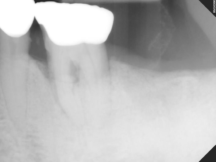
기찻길 모양의 석회화를 보이는 중막의 석회화는 고칼슘혈증 뿐만아니라 노화가 원인일 수도 있다. Figure 2는 고칼슘혈증이 없는 고령환자의 치근단방사선영상으로 하악 좌측 제1대구치의 후방 협부 연조직에 안면동맥의 석회화를 보여준다. 만일 파노라마방사선영상이나 구내방사선영상에서 이러한 동맥석회화가 관찰된다면 노화에 의한 것인지 아니면 만성신부전 또는 고칼슘혈증을 동반하는 다른 질환이 원인인지를 문진 또는 전문의 의뢰를 통해서 확인하는 절차기 필요하리라 생각된다.

